
Scientists from Nanyang Technological University, Singapore (NTU Singapore) have announced a significant breakthrough in the early detection of Alzheimer’s disease, identifying that the brain’s waste removal system often becomes obstructed long before clinical symptoms of dementia appear. The research, led by the Lee Kong Chian School of Medicine (LKCMedicine), suggests that these blockages, visible on standard brain scans, could serve as a critical early warning signal, allowing for medical intervention years before the onset of irreversible cognitive decline.
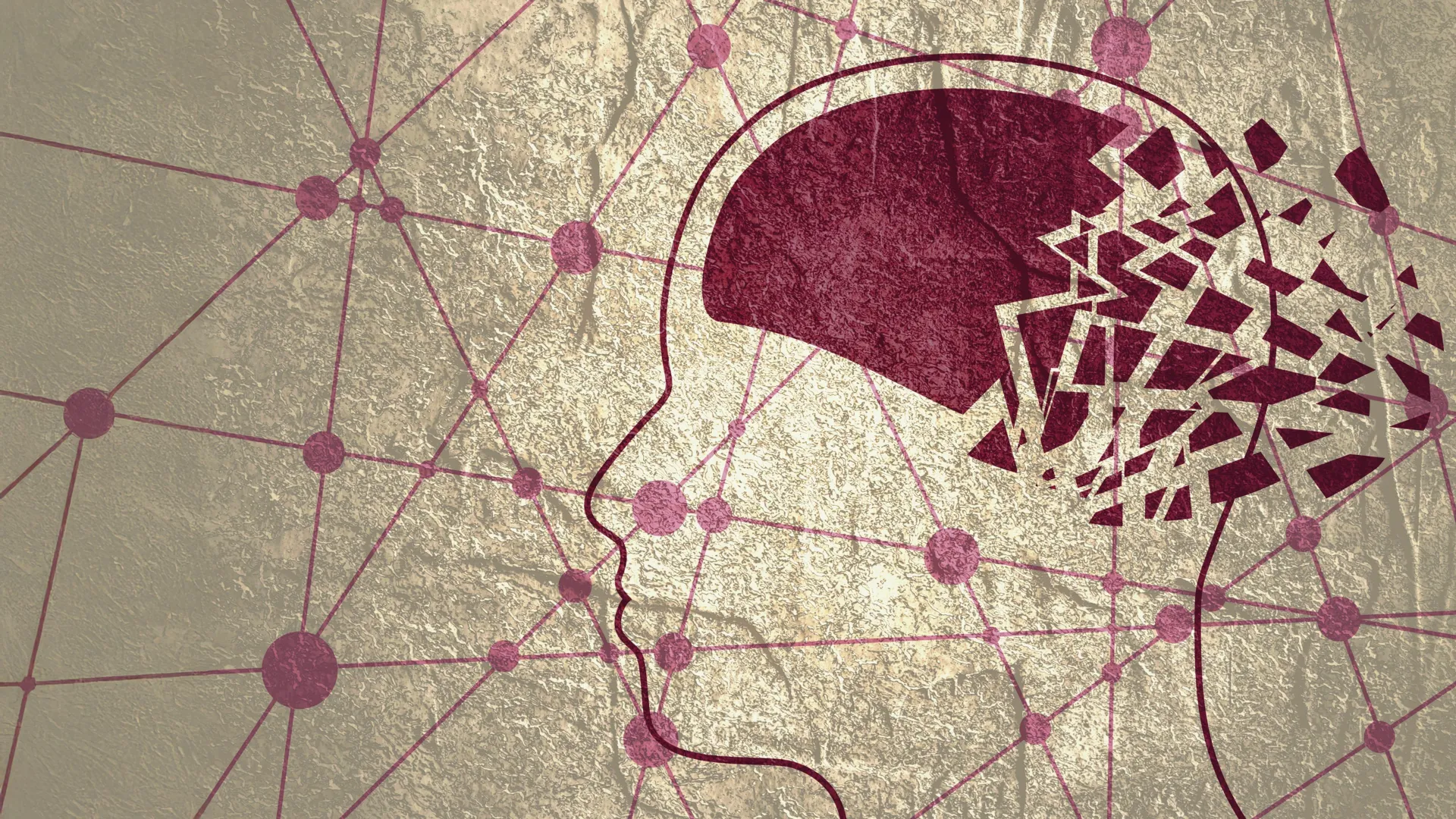
The study focuses on "enlarged perivascular spaces," which are essentially the brain’s "clogged drains." These spaces are fluid-filled channels surrounding the blood vessels in the brain that facilitate the clearance of metabolic waste. When these channels become dilated or enlarged, it indicates a failure in the brain’s glymphatic system—the network responsible for flushing out toxic proteins such as beta-amyloid and tau. The accumulation of these proteins is a hallmark of Alzheimer’s disease, leading to the eventual destruction of neurons and the loss of memory and cognitive function.
The Significance of the Asian Demographic in Neurodegenerative Research
One of the most vital aspects of this NTU Singapore study is its focus on Asian populations. Historically, the vast majority of Alzheimer’s research has been conducted on Caucasian cohorts in North America and Europe. However, dementia does not manifest identically across different ethnic and genetic backgrounds. By studying a diverse group of nearly 1,000 Singaporeans—comprising Chinese, Malay, and Indian ethnicities—the researchers have provided data that is specifically relevant to the Asian context.
Associate Professor Nagaendran Kandiah, the lead author and Director of the Dementia Research Centre (Singapore) at LKCMedicine, emphasized the genetic disparities that make regional studies essential. In Caucasian populations, the apolipoprotein E4 (APOE4) gene—a major risk factor for Alzheimer’s—is present in approximately 50 to 60 percent of patients. Conversely, in Singaporean dementia patients, this genetic marker is found in less than 20 percent of cases. This discrepancy highlights the necessity of identifying alternative biomarkers, such as enlarged perivascular spaces, that may be more indicative of disease progression in Asian communities.
The study’s findings are particularly timely given the rapidly aging populations across Asia. According to global health projections, the number of people living with dementia in the Asia-Pacific region is expected to triple by 2050. Establishing localized diagnostic criteria is therefore a matter of urgent public health priority.
Mechanisms of the Brain’s Waste Clearance System
To understand the implications of the study, it is necessary to examine how the brain maintains its health. The brain is a highly metabolic organ, producing significant amounts of waste. Unlike the rest of the body, which uses the lymphatic system to remove cellular debris, the brain utilizes perivascular spaces to circulate cerebrospinal fluid and flush out toxins.
In a healthy brain, these spaces are microscopic and efficient. However, as the brain ages or begins to develop pathology, these "drainage pipes" can become blocked. When this occurs, the spaces expand to accommodate the buildup of fluid and waste, making them visible as small, fluid-filled spots on Magnetic Resonance Imaging (MRI) scans.
The NTU research team sought to determine whether these visible enlargements were merely a sign of general aging or if they were specifically linked to the early stages of Alzheimer’s. By comparing the MRI scans of 350 individuals with normal cognitive function against those of participants with mild cognitive impairment (MCI), the team established a clear correlation. Those with MCI—a precursor to full-blown dementia—showed significantly higher rates of enlarged perivascular spaces.
Correlating Brain Scans with Blood-Based Biomarkers
The study went beyond visual imaging by incorporating advanced blood tests to measure seven specific biochemical markers associated with Alzheimer’s disease. These include various forms of beta-amyloid and tau proteins, which form the plaques and tangles that characterize the disease.
The results were definitive: enlarged perivascular spaces were strongly linked to four of the seven biochemical markers. This suggests that patients with "clogged drains" are much more likely to have high levels of toxic protein buildup in their brains. Furthermore, the researchers compared these findings to white matter damage, which has traditionally been the primary MRI marker used to assess cognitive decline.
The analysis revealed an unexpected and crucial insight: in patients with mild cognitive impairment, the presence of enlarged perivascular spaces was a more sensitive indicator of Alzheimer’s-related biochemical changes than white matter damage. This positions the waste clearance system as one of the earliest detectable markers of the disease, potentially appearing before any other structural damage to the brain is visible.
A New Perspective on the Synergy Between Vascular and Neurodegenerative Disease
For decades, the medical community viewed cerebrovascular disease (issues with blood vessels in the brain) and Alzheimer’s disease as two distinct pathways to dementia. This study challenges that binary view, demonstrating a "synergistic" relationship between the two.
Dr. Chong Yao Feng, a Consultant at the National University Hospital’s Division of Neurology, noted that the findings suggest these diseases interact in a way that accelerates decline. When the blood vessels are compromised, the waste removal system fails; when the waste removal system fails, Alzheimer’s proteins accumulate. This feedback loop suggests that treating vascular health—such as managing blood pressure and cholesterol—could have a direct impact on preventing the protein buildup associated with Alzheimer’s.
Dr. Rachel Cheong Chin Yee, a Senior Consultant at Khoo Teck Puat Hospital’s Department of Geriatric Medicine, remarked that the ability to identify high-risk individuals before symptoms appear is a "significant" advancement. It allows for a shift from reactive medicine—treating symptoms like memory loss—to proactive medicine, where doctors can focus on maintaining brain health and slowing the progression of the disease.
Clinical Implications and the Future of Diagnosis
The practical application of this research lies in its accessibility. Because perivascular spaces can be identified on routine MRI scans already used in clinical practice, there is no need for the development of expensive new hardware or invasive procedures.
Justin Ong, a fifth-year medical student at LKCMedicine and the study’s first author, highlighted that early detection gives both doctors and patients the most valuable resource: time. "Identifying Alzheimer’s sooner gives doctors more time to intervene and potentially slow the progression of symptoms such as memory loss, reduced thinking speed, and mood changes," Ong stated.
The timeline for implementing these findings into routine clinical practice could be relatively short. Since MRIs are already a standard diagnostic tool for patients reporting cognitive concerns, radiologists can be trained to specifically look for and quantify enlarged perivascular spaces as part of their standard assessment. This would provide a cost-effective way to screen for Alzheimer’s risk without the need for expensive PET scans or painful lumbar punctures.
The Path Forward: Long-Term Monitoring and Global Validation
The NTU Singapore research team is now moving into the next phase of their study, which involves a longitudinal follow-up of the participants. Over the coming years, they will track which individuals with enlarged perivascular spaces eventually progress to a clinical diagnosis of Alzheimer’s dementia. This will provide the final "gold standard" evidence needed to confirm these spaces as a reliable predictive marker.
Furthermore, the team hopes that their focus on Asian populations will inspire similar studies in other underrepresented regions. If the link between perivascular space enlargement and Alzheimer’s is validated globally across different ethnicities, it could lead to a universal standardized protocol for early dementia screening.
As the global medical community continues to search for a cure for Alzheimer’s, the consensus is shifting toward the idea that prevention and early intervention are the most effective strategies. By identifying the "clogged drains" of the brain, the scientists at NTU Singapore have provided a new lens through which we can view the earliest stages of the disease, offering hope for millions of families facing the prospect of a dementia diagnosis. This research underscores the importance of the glymphatic system in maintaining neurological health and marks a pivotal moment in the fight against one of the most challenging diseases of the 21st century.
