A landmark study from the University of California, Riverside (UCR), published in the Proceedings of the National Academy of Sciences (PNAS), provides a new framework for understanding this neurodegenerative decline. The research team, led by Seema Tiwari-Woodruff, a professor of biomedical sciences at the UCR School of Medicine, has identified mitochondrial dysfunction as a primary driver behind the death of Purkinje cells—large, specialized neurons in the cerebellum responsible for coordinating movement. The findings suggest that the metabolic "powerhouses" of these cells fail long before the cells themselves perish, offering a potential window for therapeutic intervention that could preserve mobility in MS patients.
The Biological Mechanics of Multiple Sclerosis
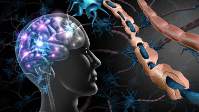
To understand the significance of the UCR study, it is necessary to examine the foundational mechanics of multiple sclerosis. MS is characterized by an aberrant immune response where the body’s own defense mechanisms attack the central nervous system. The primary target of this attack is myelin, the fatty, insulating sheath that wraps around nerve fibers (axons). Myelin functions much like the plastic coating on an electrical wire, ensuring that electrical impulses travel rapidly and efficiently between the brain and the rest of the body.
When inflammation leads to demyelination, the "insulation" is stripped away. This disruption causes electrical signals to leak, slow down, or stop entirely, resulting in the diverse array of neurological symptoms associated with MS, including vision loss, fatigue, and cognitive impairment. However, while demyelination is a hallmark of the disease, researchers have long sought to understand why the underlying neurons eventually die, a process known as neurodegeneration. The UCR study posits that the answer lies in the energy supply of the cell.
Mitochondria are organelles within cells that generate adenosine triphosphate (ATP), the chemical energy required for nearly every biological process. In the high-stakes environment of the brain, where neurons require immense amounts of energy to maintain ion gradients and fire electrical signals, mitochondrial health is paramount. The research led by graduate student Kelley Atkinson proposes that the inflammatory environment of MS creates a metabolic crisis.
"Our study proposes that inflammation and demyelination in the cerebellum disrupt mitochondrial function, contributing to nerve damage and Purkinje cell loss," explained Professor Tiwari-Woodruff. "We observed a significant loss of the mitochondrial protein COXIV in demyelinated Purkinje cells, suggesting that mitochondrial impairment contributes directly to cell death and cerebellar damage."
The Vital Role of Purkinje Neurons
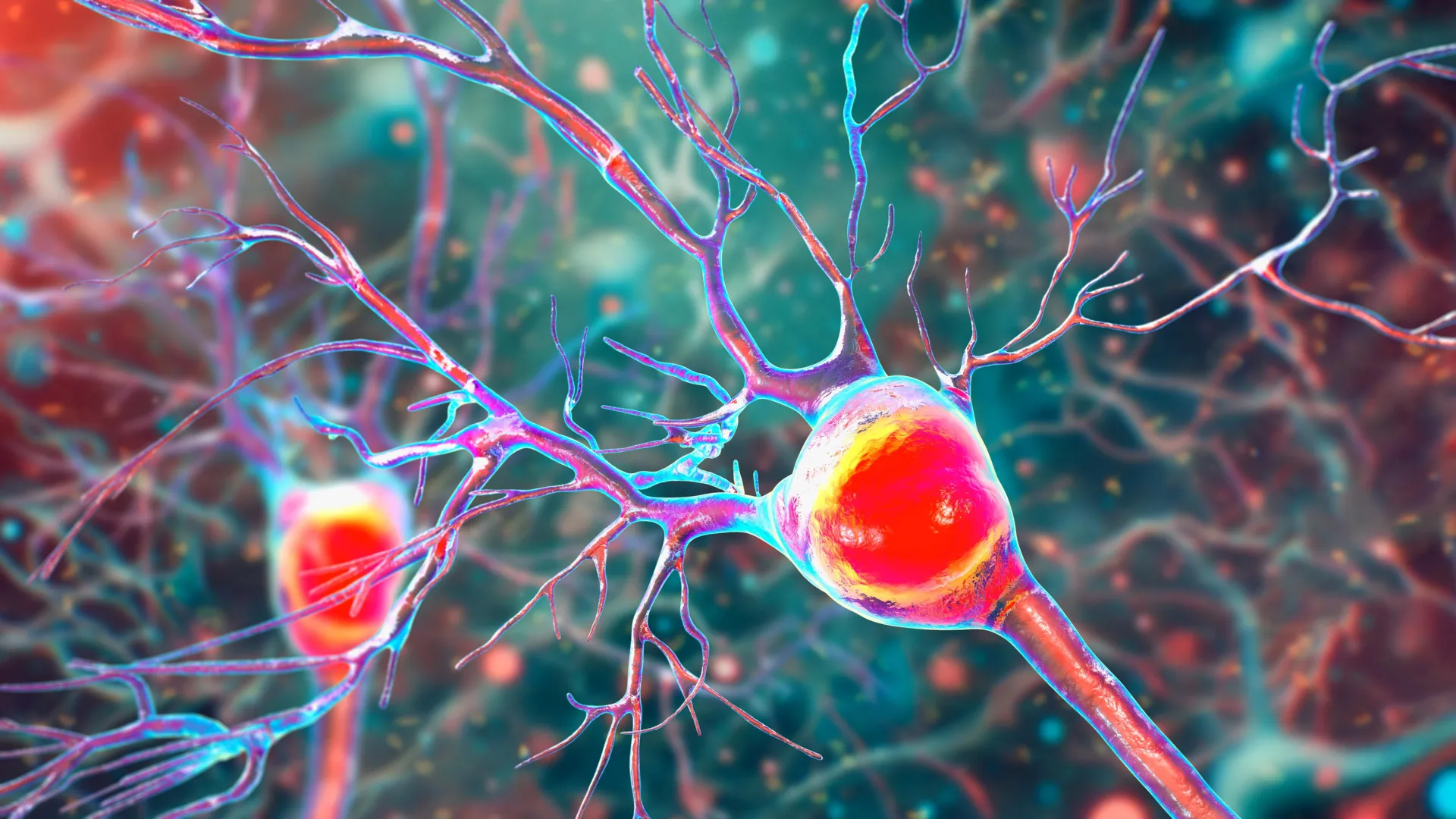
The cerebellum is often referred to as the "little brain," but its influence on human life is profound. Within its folded layers lie Purkinje cells, some of the largest and most complex neurons in the human body. These cells are characterized by their massive "dendritic trees"—elaborate branching structures that allow a single Purkinje cell to receive inputs from hundreds of thousands of other neurons.
Purkinje cells serve as the primary output for the cerebellar cortex. They are the master regulators of motor coordination, ensuring that movements are smooth, precise, and timed correctly. Whether it is the rhythmic motion of walking, the delicate finger movements of a pianist, or the reflexive adjustments required to stay upright on a moving bus, Purkinje cells are the conductors of the physiological orchestra.
In the context of MS and other neurodegenerative disorders, the loss of these cells leads to a condition known as ataxia. Patients with ataxia experience a lack of muscle control during voluntary movements, leading to a "drunken" gait, difficulty swallowing, and slurred speech. Because Purkinje cells are so metabolically active, they are particularly vulnerable to any disruption in their energy supply.
"Inside the cerebellum are special cells called Purkinje neurons," Tiwari-Woodruff noted. "These large, highly active cells help coordinate smooth, precise movements. They’re essential for balance and fine motor skills. As these neurons disappear, people may develop ataxia. Understanding why they’re damaged in MS could help us find better treatments to protect movement and balance."
Chronology of Decay: From Inflammation to Energy Failure
The UCR researchers utilized a dual-track approach to investigate the progression of cerebellar damage, analyzing both human postmortem tissue and animal models. By examining cerebellar tissue from individuals who had secondary progressive MS—a stage of the disease where disability increases steadily—and comparing it with tissue from healthy donors, the team was able to map the physical manifestations of the disease.
The human tissue samples, provided by the National Institutes of Health’s NeuroBioBank and the Cleveland Clinic, revealed a stark reality: Purkinje cells in MS-affected brains had significantly fewer branches (dendrites), showed evidence of advanced demyelination, and lacked critical mitochondrial proteins.
To observe how these changes unfold in real-time, the team employed the Experimental Autoimmune Encephalomyelitis (EAE) mouse model. EAE is a widely accepted surrogate for MS in research, as it mimics the immune-mediated demyelination and subsequent neurodegeneration seen in humans. The chronology of the disease in the mouse model provided a clear timeline:
- Early Phase: Myelin breakdown begins shortly after the onset of the disease. While the neurons remain intact, their protective coating is compromised.
- Intermediate Phase: Mitochondrial proteins, specifically COXIV, begin to diminish. This marks the onset of the "energy crisis," where the cell can no longer produce enough ATP to sustain its complex structure.
- Late Phase: The Purkinje cells, starved of energy and stripped of their insulation, undergo apoptosis (programmed cell death). This is the point where cerebellar volume is lost and motor deficits become permanent.
"The remaining neurons don’t work as well because their mitochondria start to fail," Tiwari-Woodruff said. "These problems—less energy, loss of myelin, and damaged neurons—start early, but the actual death of the brain cells tends to happen later. The loss of energy in brain cells seems to be a key part of what causes damage in MS."
Supporting Data and Statistical Significance
The study’s findings are bolstered by quantitative data regarding the loss of COXIV, a subunit of the cytochrome c oxidase enzyme. This enzyme is a critical component of the mitochondrial electron transport chain, which is responsible for the final steps of ATP production.
In the demyelinated regions of the MS cerebellum, the researchers found a statistically significant reduction in COXIV expression compared to myelinated regions and healthy controls. This correlation suggests that the loss of myelin and the presence of inflammatory cytokines (signaling molecules) create a toxic environment that specifically targets mitochondrial integrity.
Furthermore, the study highlighted a "dying-back" phenomenon in Purkinje cells. This occurs when the furthest reaches of the neuron—the dendritic branches—begin to wither away before the main cell body dies. This structural atrophy directly correlates with the severity of motor impairment observed in the EAE models, providing a physical metric for the progression of disability.
Broader Implications for MS Therapeutics
The identification of mitochondrial failure as a central player in MS neurodegeneration shifts the focus of potential treatments. Currently, most MS therapies are "immunomodulatory," meaning they work by suppressing or altering the immune system to prevent new inflammatory attacks. While these drugs are effective at reducing the frequency of relapses in early-stage MS, they have limited success in stopping the slow, "smoldering" progression of the disease once it has taken hold in the cerebellum.
The UCR research suggests a "neuroprotective" strategy. If clinicians can bolster mitochondrial health or stabilize energy production in Purkinje cells, they might be able to keep those cells alive even in the face of ongoing inflammation.
"Targeting mitochondrial health may represent a promising strategy to slow or prevent neurological decline and improve quality of life for people living with MS," Tiwari-Woodruff stated. This could involve the development of drugs that enhance mitochondrial biogenesis (the creation of new mitochondria), antioxidants that protect mitochondrial membranes from oxidative stress, or metabolic supplements that provide alternative energy sources for stressed neurons.
Future Research and the Role of Glial Cells
The UCR team is not stopping at Purkinje cells. The brain is a complex ecosystem where neurons rely on "support cells" known as glia. The next phase of their research involves investigating whether mitochondrial damage extends to:
- Oligodendrocytes: The cells responsible for creating myelin. If their mitochondria fail, they cannot repair the myelin sheath, leading to permanent demyelination.
- Astrocytes: Star-shaped cells that provide nutrients to neurons and maintain the blood-brain barrier. Malfunctioning astrocytes can exacerbate inflammation and fail to clear toxic metabolic byproducts.
"To answer this, one of our ongoing research projects is focused on studying mitochondria in specific types of brain cells in the cerebellum," Tiwari-Woodruff said. "Such research can open the door to finding ways to protect the brain early on—like boosting energy in brain cells, helping them repair their protective myelin coating, or calming the immune system before too much damage is done."
The Call for Continued Scientific Investment
The study, funded by the National Multiple Sclerosis Society, underscores the necessity of long-term investment in basic and clinical science. As the global population ages and the prevalence of MS continues to rise in certain regions, the economic and social burden of the disease grows.
Tiwari-Woodruff emphasized that breakthroughs of this nature are only possible through sustained public and private support. "Cutting funding to science only slows progress when we need it most," she said. "Public support for research matters now more than ever."
By pinpointing the exact metabolic failures that lead to the loss of balance and coordination, the UCR study provides a roadmap for the next generation of MS treatments. For the millions of people living with the fear of losing their mobility, this research offers more than just data; it offers a path toward a future where the progressive decline of MS is no longer an inevitability.