
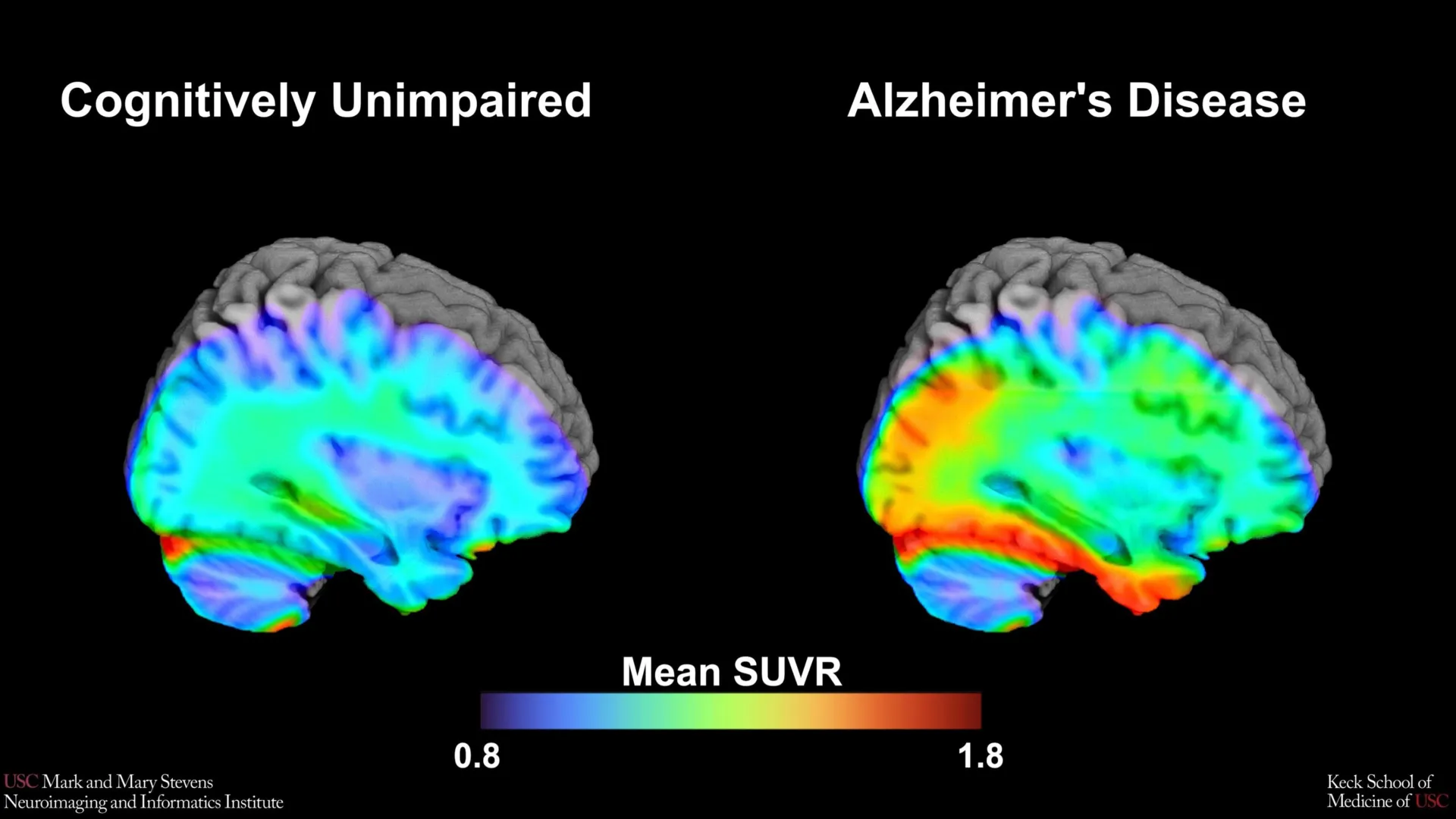
The research was conducted as part of the Health and Aging Brain Study-Health Disparities (HABS-HD), a comprehensive, multi-university collaboration led by the University of North Texas Health Science Center. Supported by the National Institute on Aging, the HABS-HD initiative represents one of the most ambitious efforts to date to study Alzheimer’s disease and related dementias within a framework of health equity, moving beyond the traditionally narrow demographics of clinical trials.
The Science of Tau PET Imaging and the 18F-PI-2620 Tracer
To understand the significance of this benchmark, it is essential to examine the underlying pathology of Alzheimer’s disease. For decades, researchers focused primarily on amyloid-beta plaques—sticky protein fragments that build up between nerve cells. However, more recent research has shifted focus toward tau, a protein that forms "tangles" inside the neurons themselves. While amyloid is often seen as an early indicator of the disease process, tau tangles are much more closely correlated with the actual onset of memory loss and cognitive decline.
The USC team utilized a sophisticated imaging technique known as tau Positron Emission Tomography (PET). This process involves injecting a patient with a specialized radioactive tracer that binds to tau proteins in the brain. For this study, the researchers used a second-generation tracer called 18F-PI-2620. This particular tracer is noted for its high affinity and selectivity, allowing researchers to visualize tau accumulation with greater precision than earlier imaging agents.
By analyzing the scans of over 675 older adults, the researchers sought to establish "tau cut-points." A cut-point is a numerical threshold of protein density. If a patient’s scan shows tau levels above this threshold, it serves as a biological red flag, suggesting that the individual is experiencing clinically relevant changes associated with Alzheimer’s rather than normal age-related decline.
Chronology of the Study and Findings
The study began with the recruitment of a diverse cohort of participants through the HABS-HD framework. Participants underwent a battery of cognitive tests to determine their level of impairment, ranging from those with normal cognitive function to those with Mild Cognitive Impairment (MCI) or early-stage Alzheimer’s. Following these assessments, the participants underwent PET imaging to measure both amyloid and tau levels.
The researchers focused their analysis on the medial temporal lobe, a region deep within the brain that serves as the hub for memory and navigation. This area is typically the first to show signs of tau accumulation in the early stages of Alzheimer’s.
Upon analyzing the data, a clear pattern emerged for Hispanic and non-Hispanic White participants. In these groups, when tau levels in the medial temporal lobe exceeded the established cut-point, there was a strong correlation with cognitive impairment—but primarily when amyloid plaques were also present. This "dual-protein" requirement aligns with the current biological definition of Alzheimer’s disease as a "proteinopathy" involving both amyloid and tau.
However, the results for non-Hispanic Black participants presented a stark contrast. In this group, the established tau cut-point failed to reliably distinguish between those with cognitive impairment and those without. This finding suggests that the traditional "amyloid-tau" model of Alzheimer’s may not tell the whole story for Black Americans, indicating that other factors—such as vascular health, chronic inflammation, or social determinants of health—might be the primary drivers of cognitive decline in this demographic.
Addressing the Diversity Gap in Alzheimer’s Research
The disparity in the study’s results highlights a long-standing issue in neurology: the lack of diversity in clinical research. Historically, the vast majority of Alzheimer’s data has been gathered from non-Hispanic White populations. This has led to a "one-size-fits-all" diagnostic approach that may not be accurate for minority groups, who often face higher rates of dementia.
"Our study is an important step toward better understanding how tau relates to cognition in diverse populations and has important implications for future clinical trials that aim to target tau," said senior author Meredith N. Braskie, PhD, an assistant professor of neurology at USC. Dr. Braskie noted that the failure of the tau cut-point in Black participants suggests that the medical community must look beyond a single protein to understand brain health.
Lead author Victoria R. Tennant, a PhD candidate in USC’s Neuroscience Graduate Program, echoed this sentiment. She emphasized that while the medial temporal lobe remains a critical area for study, the limited reliability of tau in certain groups necessitates a broader perspective. "The findings highlight the need for more diverse populations in research and for future studies to examine both biological and social determinants of Alzheimer’s disease," Tennant stated.
Supporting Data and Regional Specificity
The data produced by the USC team provides a roadmap for how clinicians might interpret brain scans in the future. By focusing on the medial temporal lobe, the study identified that this specific region acts as a "canary in the coal mine" for many patients.
Key statistical takeaways from the study include:
- Participant Volume: 675+ individuals, making it one of the largest tau PET studies focused on health disparities.
- Tracer Efficacy: 18F-PI-2620 demonstrated a high signal-to-noise ratio, making the identification of the cut-point mathematically robust for the groups where it was effective.
- The Amyloid Link: The correlation between tau and cognitive impairment was significantly stronger in the presence of amyloid, reinforcing the theory that these two proteins work in tandem to degrade neurological function.
This regional specificity is vital. By knowing exactly where to look (the medial temporal lobe) and what threshold to look for, clinicians can move away from subjective assessments and toward objective, data-driven diagnoses.
Institutional Perspectives and Broader Impact
The Mark and Mary Stevens Neuroimaging and Informatics Institute (Stevens INI) has long been at the forefront of mapping the human brain. For Director Arthur W. Toga, PhD, this study is a testament to the power of large-scale, collaborative data.
"This type of imaging is critical for understanding who is at risk and how the disease develops," Toga said. He highlighted that the HABS-HD project has already yielded significant insights into vascular contributions to dementia and the role of socioeconomic status in cognitive aging. "We hope this work will lead to more personalized care and better outcomes for all communities," he added.
The implications for clinical trials are particularly profound. Currently, many pharmaceutical companies are developing "tau-targeting" drugs designed to clear these tangles from the brain. If a trial uses a tau cut-point based solely on White populations to recruit participants, it may inadvertently exclude or misdiagnose Black or Hispanic patients, leading to skewed results and treatments that are not effective for the general population.
Analysis of Implications: Toward Personalized Neurology
The USC study marks a transition from a generalized understanding of Alzheimer’s to a more nuanced, personalized approach. If the drivers of cognitive decline differ across racial and ethnic lines, then the "cure" for Alzheimer’s may not be a single drug, but a suite of interventions tailored to an individual’s specific biological and social profile.
For Hispanic and White populations, the tau cut-point in the medial temporal lobe offers a promising diagnostic benchmark that could allow for earlier intervention. Early detection is crucial, as many of the newest Alzheimer’s treatments are most effective when administered before significant brain damage has occurred.
For non-Hispanic Black populations, the study’s results are a call to action. They suggest that researchers must investigate "mixed pathologies"—the combination of Alzheimer’s changes with other conditions like small-vessel disease or hypertension-related brain injury. By identifying the unique biomarkers for these groups, the medical community can ensure that no population is left behind in the race to end dementia.
Conclusion and Future Directions
The research conducted by the Stevens INI team, in collaboration with the HABS-HD partners, provides a critical update to the toolkit available to neurologists. While the identification of a tau cut-point using the 18F-PI-2620 tracer is a major technical achievement, the study’s most enduring legacy may be its contribution to health equity.
As the global population ages, the burden of Alzheimer’s disease is expected to triple by 2050. Success in managing this crisis will depend on the ability of researchers to develop diagnostic tools that are as diverse as the patients they serve. The work at USC demonstrates that while the brain is a universal organ, the paths it takes toward disease are deeply influenced by a complex interplay of genetics, environment, and biology. Moving forward, the focus will remain on refining these benchmarks and exploring the "other pathologies" that drive cognitive decline, ensuring that personalized, effective care becomes a reality for all.