A groundbreaking study led by researchers at the University of California, Riverside (UCR), recently published in the Proceedings of the National Academy of Sciences (PNAS), has identified a critical driver of this neurodegenerative process. The research points toward malfunctioning mitochondria—the intracellular structures responsible for energy production—as the primary culprit behind the death of Purkinje cells. These large, specialized neurons are the sole output of the cerebellar cortex, and their loss is directly correlated with the worsening physical impairment observed in patients with progressive forms of MS.
The Triad of Destruction: Inflammation, Demyelination, and Energy Failure
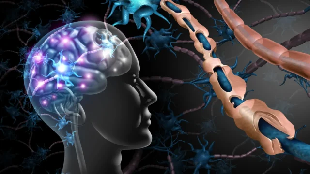
The traditional understanding of multiple sclerosis centers on the immune system’s misguided attack on myelin, the fatty insulation that wraps around nerve fibers (axons). This process, known as demyelination, disrupts the rapid transmission of electrical impulses, effectively "short-circuiting" the brain’s communication network. However, the UCR study suggests that demyelination is only one part of a more complex cascade of cellular failure.
Mitochondria serve as the "powerhouses" of the cell, converting nutrients into adenosine triphosphate (ATP), the chemical energy that fuels every cellular process. In the context of MS, the UCR research team, led by Professor Seema Tiwari-Woodruff and graduate student Kelley Atkinson, discovered that the inflammatory environment of the cerebellum induces a state of chronic metabolic stress.
"Our study proposes that inflammation and demyelination in the cerebellum disrupt mitochondrial function, contributing to nerve damage and Purkinje cell loss," explained Dr. Tiwari-Woodruff, a professor of biomedical sciences at the UC Riverside School of Medicine. The researchers identified a specific deficit in a mitochondrial protein known as COXIV. This protein is essential for the electron transport chain, the final stage of energy production. When COXIV levels drop, the mitochondria can no longer meet the high energy demands of the cell, leading to a state of "energy failure" that eventually triggers programmed cell death.
The Critical Role of Purkinje Neurons in Human Mobility
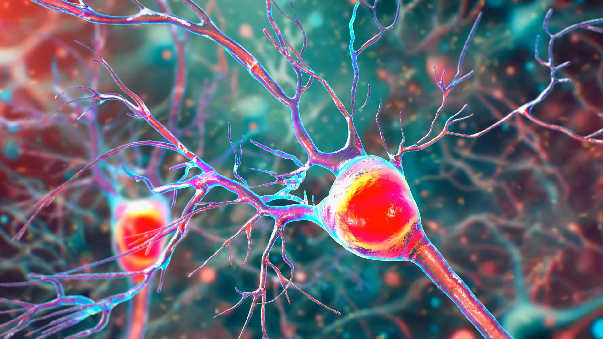
To understand the gravity of this discovery, one must look at the unique role of Purkinje cells within the human body. These neurons are among the largest in the brain and possess intricate, tree-like dendritic structures that allow them to receive and process a massive amount of sensory information. They act as the "precision engineers" of movement, synthesizing data from the inner ear, the eyes, and the muscles to ensure that every step, reach, or gesture is smooth and coordinated.
When these cells are healthy, they facilitate complex tasks such as dancing, typing, or maintaining balance on an uneven surface. However, in the presence of MS-related damage, these neurons begin to lose their dendritic branches—a process akin to a tree losing its limbs. This structural atrophy occurs alongside the loss of myelin, leaving the neuron vulnerable.
"Inside the cerebellum are special cells called Purkinje neurons," Tiwari-Woodruff stated. "They’re essential for balance and fine motor skills. Because Purkinje cells play such a central role in movement, their loss can cause serious mobility issues. Understanding why they’re damaged in MS could help us find better treatments to protect movement and balance in people with the disease."
The loss of these neurons leads to ataxia, a neurological sign consisting of a lack of voluntary coordination of muscle movements. For many MS patients, ataxia is one of the most life-altering aspects of the disease, as it can eventually lead to the loss of independence and the requirement of mobility aids.
Insights from the EAE Mouse Model and Human Pathology
The research team utilized a multi-faceted approach to reach their conclusions, combining the analysis of human tissue with longitudinal studies of animal models. They examined postmortem cerebellar tissue from individuals who had been diagnosed with secondary progressive MS—a stage of the disease characterized by a steady worsening of symptoms without periods of remission. This tissue was compared against samples from healthy donors, provided by the National Institutes of Health’s NeuroBioBank and the Cleveland Clinic.
The human samples revealed a stark reality: Purkinje cells in the MS-affected brains were significantly reduced in number, and those that remained showed clear signs of mitochondrial dysfunction and structural decay.
To observe how these changes unfold in real-time, the researchers employed the experimental autoimmune encephalomyelitis (EAE) mouse model. EAE is a widely accepted scientific proxy for MS, as it mimics the autoimmune-driven demyelination and neurodegeneration seen in humans. By tracking the mice as the disease progressed, the team was able to establish a timeline of destruction.
The data showed that myelin breakdown occurs early in the disease course, followed closely by a decline in mitochondrial activity. While the neurons initially survive these metabolic insults, they eventually reach a breaking point. The actual death of the brain cells tends to occur in the later, more severe stages of the disease, suggesting a "window of opportunity" where intervention might be possible before the damage becomes irreversible.
"The loss of energy in brain cells seems to be a key part of what causes damage in MS," Tiwari-Woodruff noted. This finding shifts the focus of potential therapies from simply suppressing the immune system to actively protecting the metabolic health of the neurons.
Implications for Future Therapeutic Strategies
The identification of mitochondrial failure as a primary driver of Purkinje cell loss opens a new frontier for MS treatment. Most current MS medications are immunomodulators, designed to reduce the frequency of relapses by preventing immune cells from entering the central nervous system. While effective for relapsing-remitting MS (RRMS), these drugs often fail to stop the slow, "smoldering" progression of disability seen in secondary progressive MS (SPMS) and primary progressive MS (PPMS).
The UCR findings suggest that a dual-action approach may be necessary: one that calms the immune system while simultaneously boosting mitochondrial health. Potential treatments could include neuroprotective agents that stabilize the COXIV protein, antioxidants that reduce the oxidative stress caused by failing mitochondria, or metabolic stimulants that help neurons maintain ATP production despite the loss of myelin.
"Targeting mitochondrial health may represent a promising strategy to slow or prevent neurological decline and improve quality of life for people living with MS," said Tiwari-Woodruff. This research brings the scientific community a step closer to "neuroprotective" medicine—a long-sought goal in the treatment of all neurodegenerative diseases, including Parkinson’s and Alzheimer’s.
Beyond Purkinje Cells: A Comprehensive View of the Cerebellum
The UCR research team is not stopping at Purkinje cells. Their ongoing projects are investigating whether mitochondrial damage extends to other critical cell types within the cerebellum, such as:
- Oligodendrocytes: The cells responsible for producing and repairing the myelin sheath. If their mitochondria fail, they cannot repair the damage caused by MS attacks.
- Astrocytes: Star-shaped cells that provide structural support and nutrients to neurons. Malfunctioning astrocytes can exacerbate inflammation and leave neurons without the support they need to survive.
By understanding the mitochondrial health of the entire cerebellar ecosystem, researchers hope to develop a holistic "repair kit" for the brain. This would involve helping cells repair their protective coatings while ensuring they have the energy required to maintain their complex electrical signaling.
The Socio-Economic Context and the Call for Continued Research
The study, supported by the National Multiple Sclerosis Society, highlights the vital importance of sustained investment in biomedical research. As the global population ages and the prevalence of MS continues to rise in certain regions, the economic and social burden of the disease grows. The cost of MS, including medical care and lost productivity, is estimated to be in the billions of dollars annually in the United States alone.
Professor Tiwari-Woodruff emphasized that scientific progress is a marathon, not a sprint, and requires consistent public and private support. "Cutting funding to science only slows progress when we need it most," she remarked. "Public support for research matters now more than ever."
The research team, which included Shane Desfor, Micah Feria, Maria T. Sekyia, Marvellous Osunde, Sandhya Sriram, Saima Noori, Wendy Rincón, and Britany Belloa, has provided a new roadmap for understanding the "hidden" damage of MS. By focusing on the microscopic power plants within our brain cells, science may finally find a way to preserve the movement, balance, and dignity of those living with this challenging condition.
As the scientific community moves forward, the UCR study will serve as a foundational piece of evidence in the shift toward metabolic-based neuroprotection. The ultimate goal remains clear: to transform MS from a progressive, debilitating disease into a manageable condition where the brain’s ability to heal and power itself is never lost.