
The Evolving Landscape of NPC Treatment
Nasopharyngeal carcinoma (NPC), a prevalent epithelial malignancy originating from the nasopharyngeal mucosal lining, has historically posed significant treatment challenges. Its deep anatomical location within the nasopharynx and a high propensity for lymph node metastasis have traditionally necessitated aggressive treatment modalities, with radiotherapy being the standard of care, even for early-stage presentations. However, advancements in public health education and sophisticated early cancer screening programs have led to a notable increase in the detection of NPC at its nascent stages. Studies indicate that a substantial proportion of NPC patients, ranging from 47.1% to 51.1%, are diagnosed with Stage I disease, characterized by lesions confined to the nasopharyngeal cavity, within screening populations. This demographic shift has underscored an urgent need for therapeutic strategies that are less invasive than radiotherapy, paving the way for the exploration of endoscopic surgical interventions.
Prior research has already demonstrated the feasibility of endoscopic nasopharyngectomy (ENPG) for the resection of newly diagnosed Stage I NPCs, with subsequent studies identifying specific patient cohorts who could benefit from such procedures. A prospective clinical trial (NCT03353467) is currently underway to rigorously evaluate the efficacy and safety of endoscopic surgery for Stage I NPC. Nevertheless, existing ENPG techniques often rely heavily on the surgeon’s intricate anatomical knowledge and surgical dexterity, particularly when tumors are situated in challenging areas like the pharyngeal recess and the lateral wall of the nasopharynx.
To address these complexities, a series of innovative techniques have been developed and implemented. These include the "third-hand technique," "cube-like resection," "dumpling-making technique," and the application of a "pedicle nasal mucoperiosteal flap." These advancements aim to facilitate en bloc resection, protect the critical internal carotid artery (ICA), and effectively reconstruct the resulting surgical defects. Despite these innovations, the pursuit of an improved surgical perspective, enhanced anatomical clarity, and more standardized surgical protocols remains paramount to achieve a delicate balance between radical tumor removal and minimizing the risk of ICA injury.
Development of the NESD Technique
Years of dedicated effort and refinement have culminated in the development of the Nasopharyngeal Endoscopic Submucosal Dissection (NESD) technique. This novel approach is designed to enable the safe and complete resection of the entire nasopharyngeal mucosa, encompassing the pharyngeal recess and lateral wall, while providing optimal protection for the internal carotid artery. This report marks the first publication detailing the initial surgical experience utilizing the NESD technique for localized early-stage NPC.
Case Study: A Pioneer in NESD
The efficacy of the NESD technique was recently showcased in the case of a 40-year-old male patient who presented with an incidentally discovered neoplasm on the right and posterior nasopharyngeal wall during a routine health examination. Subsequent pathological analysis confirmed the lesion to be an undifferentiated nonkeratinizing carcinoma. Comprehensive diagnostic evaluations, including magnetic resonance imaging (MRI), positron emission tomography-computed tomography (PET-CT), and endoscopic examination, were performed to meticulously assess the extent of tumor invasion. The patient was ultimately diagnosed with T1N0M0 Stage I NPC, adhering to the eighth edition of the Union for International Cancer Control (UICC) staging system. Following a thorough discussion of treatment options, the patient opted for the NESD procedure, electing to forgo radiotherapy.
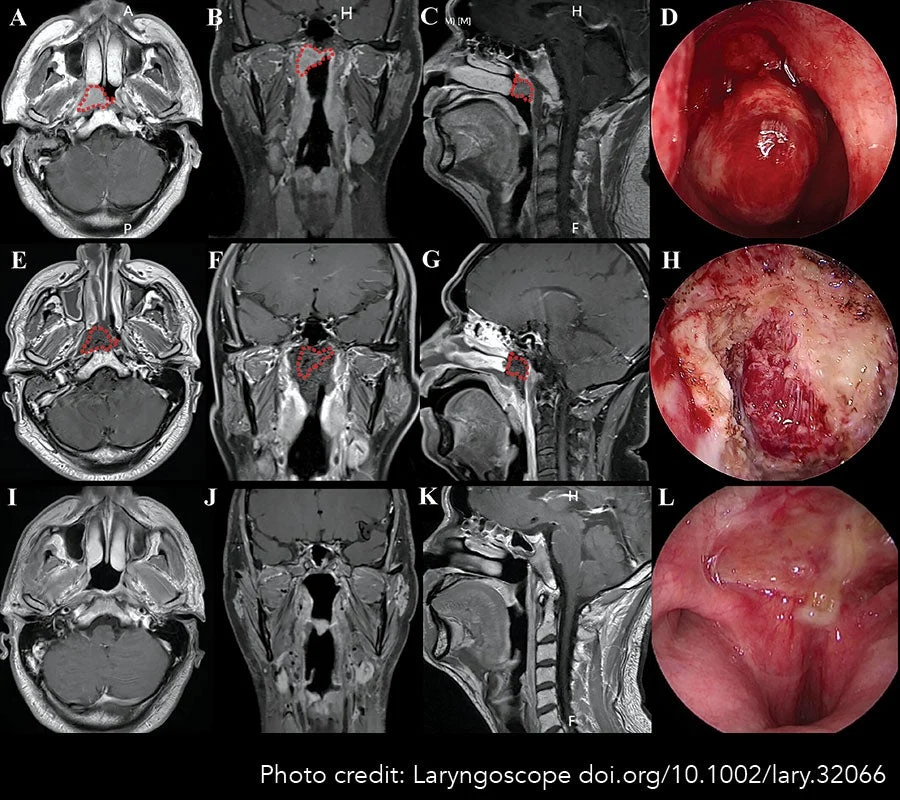
Pre-operative planning meticulously defined the surgical margins, encompassing the entirety of the nasopharyngeal mucosa, based on detailed MRI assessments (Figure 1A-C). The study protocol received approval from the Clinical Research Ethics Committee of the Fifth Affiliated Hospital of Guangzhou Medical University, and the patient provided written informed consent for participation.

The NESD Surgical Procedure: A Step-by-Step Breakdown
The NESD procedure was performed under general anesthesia. Following standard head and neck preparation and draping, nasal endoscopic guidance was used to administer a solution of 1:10,000 epinephrine to induce shrinkage of the nasal concha.
Imaging of the nasopharyngeal cavity revealed its approximate "cube" shape. The identified tumor was located on the right and posterior walls of the nasopharynx (Figure 2A). The NESD procedure, guided by a 4-mm rigid endoscope via an oronasal approach, involved a meticulously orchestrated seven-step process for the complete removal of the nasopharyngeal mucosa.
Step One: Delineation of Resection Boundaries: The initial step involved marking the planned resection boundary using electrocoagulation, precisely defining the surgical incisions around the nasopharyngeal cavity.
Step Two: Mobilization of the Anterior Wall: The anterior wall was freed by dividing the posterior mucosa of the nasal septum. The vomer bone was then polished to enhance the operative space within the nasopharynx (Figure 2B).
Step Three: Excision of the Superior Wall: The entire superior wall of the nasopharynx was carefully separated from the upper margin of the posterior nostril, following the bony surface all the way to the clivus (Figure 2C).
Step Four: Establishment of the Trans-Oral-Nasal Approach (TONA): A mouth gag was employed to ensure the patient’s mouth remained open. Two catheters were inserted through the bilateral nasal cavities and the oropharyngeal cavity, with their tips being retrieved from the oral cavity. These catheters served to suspend the soft palate. This combined TONA approach, integrating oral, oropharyngeal, and nasal pathways, significantly broadens the surgical perspective, creates a more spacious operating environment, and facilitates more convenient surgical access (Figure 2D).
Step Five: Separation of the Posterior Wall: A crucial "safety tunnel" was meticulously established to ensure a secure operative space. The mucosa of the posterior nasopharyngeal wall, at the level of the cephalopharyngeus, was incised and carefully dissected along the surface of the longus capitis muscle, extending up to the pharyngeal raphe. Throughout this dissection, the integrity of the membrane of the longus capitis muscle was rigorously maintained. Subsequently, the loose connective tissue space within the submucosa, situated at the junction of the posterior and lateral nasopharyngeal mucosal walls, was meticulously separated. This dissection extended towards the lower region of the apex of the partis petrous ossis temporalis and the inner aspect of the external orifice of the ICA sheath, thereby establishing a safe tunnel that effectively separated the nasopharyngeal mucosa from the vicinity of the ICA. The connective tissue was then excised inward and upward until it met the superior wall incision (Figure 2E).
Step Six: Resection of the Left Wall: The mucosa at the mid-point of the left Eustachian tube carina was incised. The mucosal tissue was then separated along the submucosal space towards the parapharyngeal space, and the pharynx was recessed until the deepest corner of the established safety tunnel was reached (Figure 2F). The incisions were subsequently converged with the nasopharyngeal superior and posterior wall incisions to achieve a complete resection of the left nasopharyngeal wall (Figure 2G). Extreme caution was exercised to avoid exceeding the boundaries of the safety tunnel, thereby preventing injury to the parapharyngeal vessels and nerves.

Step Seven: Resection of the Right Wall: A right-sided safety tunnel was created using the identical technique employed for the left tunnel (Figure 2H). Subsequently, the Eustachian tube carina mucosa was divided and separated until the right recess was reached through the submucosal space. This dissection converged with the contralateral nasopharyngeal superior and posterior wall incisions along the surface of the longus capitis muscle (Figure 2I), ensuring the complete dissociation of the entire nasopharyngeal mucosa from the tumor.
The gross specimen, comprising the tumor enveloped by the entire nasopharyngeal mucosa, was finally removed orally. This en bloc resection ensured sufficient safe margins around the tumor, confirming the complete removal of the total nasopharyngeal mucosa (Figure 2J-L).
Following the primary resection, meticulous hemostasis was achieved. The operative cavity was irrigated with distilled water to mitigate the risk of tumor seeding. Multiple biopsies were taken from the margins of the incision for immediate pathological examination. A bioresorbable membrane was then applied to cover the nasopharyngeal wound, promoting reepithelialization. A gelatin sponge and absorbable fibrin glue were utilized to fill the operative cavity. Upon completion of the procedure, no active bleeding was observed in the oropharynx or bilateral nostrils.
Post-Operative Care and Follow-Up Protocol
Post-operative management involved regular endoscopic assessments of wound healing every two weeks until complete reepithelialization was achieved. Subsequent follow-up appointments were scheduled at three-month intervals for the first three years, at least every six months for years four and five, and annually thereafter. Each assessment included nasopharyngoscopy, head and neck MRI, and quantification of Epstein-Barr virus (EBV)-DNA copy numbers. Conventional examinations, such as chest X-rays and abdominal ultrasounds, were conducted every six to twelve months. If deemed necessary, more comprehensive whole-body examinations, including skeletal scintigraphy or PET/CT scans, were performed.
Remarkable Outcomes and Patient Recovery
The surgical procedure was completed within a duration of 130 minutes, with an estimated blood loss of 50 mL. The patient reported no post-operative complications, such as headaches or nosebleeds. Post-operative MRI scans performed within the first week confirmed the complete resection of the primary tumor and the entire nasopharyngeal submucosa (Figure 1E-H).
Histopathological examination of the margin biopsies revealed no evidence of residual tumor. The surgical wound achieved complete healing within three weeks post-operation. Over a two-year follow-up period, the patient experienced no tumor recurrence or development of sequelae commonly associated with NPC treatment, including dry mouth, hearing impairment, otitis, trismus, dysphagia, or nasal obstruction (Figure 1I-L).
Implications and Future Directions
The successful application of the NESD technique in this early case study offers a compelling glimpse into a future where early-stage NPC can be managed with greater precision and reduced morbidity. By enabling the complete resection of the nasopharyngeal mucosa in a minimally invasive manner, NESD holds the potential to significantly improve the quality of life for patients by sparing them the long-term side effects of radiotherapy, such as xerostomia, osteoradionecrosis, and potential secondary malignancies.
The ongoing prospective study (NCT03353467) will be crucial in further validating the long-term efficacy and safety of endoscopic surgery for Stage I NPC. As the NESD technique continues to be refined and more surgeons gain proficiency, it is poised to become a cornerstone of treatment for select early-stage NPC patients, offering a more personalized and less burdensome approach to cancer care. This development represents a significant stride forward in the management of nasopharyngeal carcinoma, aligning with the broader medical trend towards less invasive and more patient-centered oncological treatments.