The Biological Mechanism of Cerebellar Decline
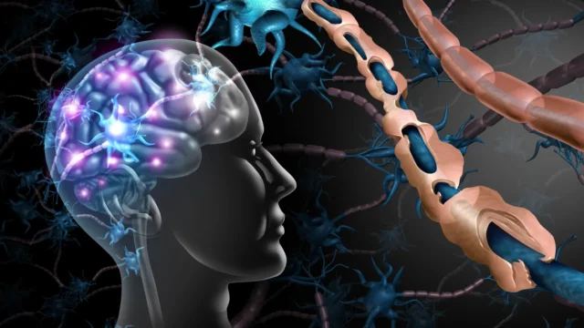
Multiple sclerosis is pathologically defined by chronic inflammation and demyelination. The myelin sheath acts as an insulating layer for axons, much like the plastic coating on an electrical wire. When this insulation is stripped away through an autoimmune response, the electrical impulses traveling between the brain and the rest of the body are slowed or blocked entirely. However, the UCR study suggests that the loss of myelin is only the beginning of a cascading failure.
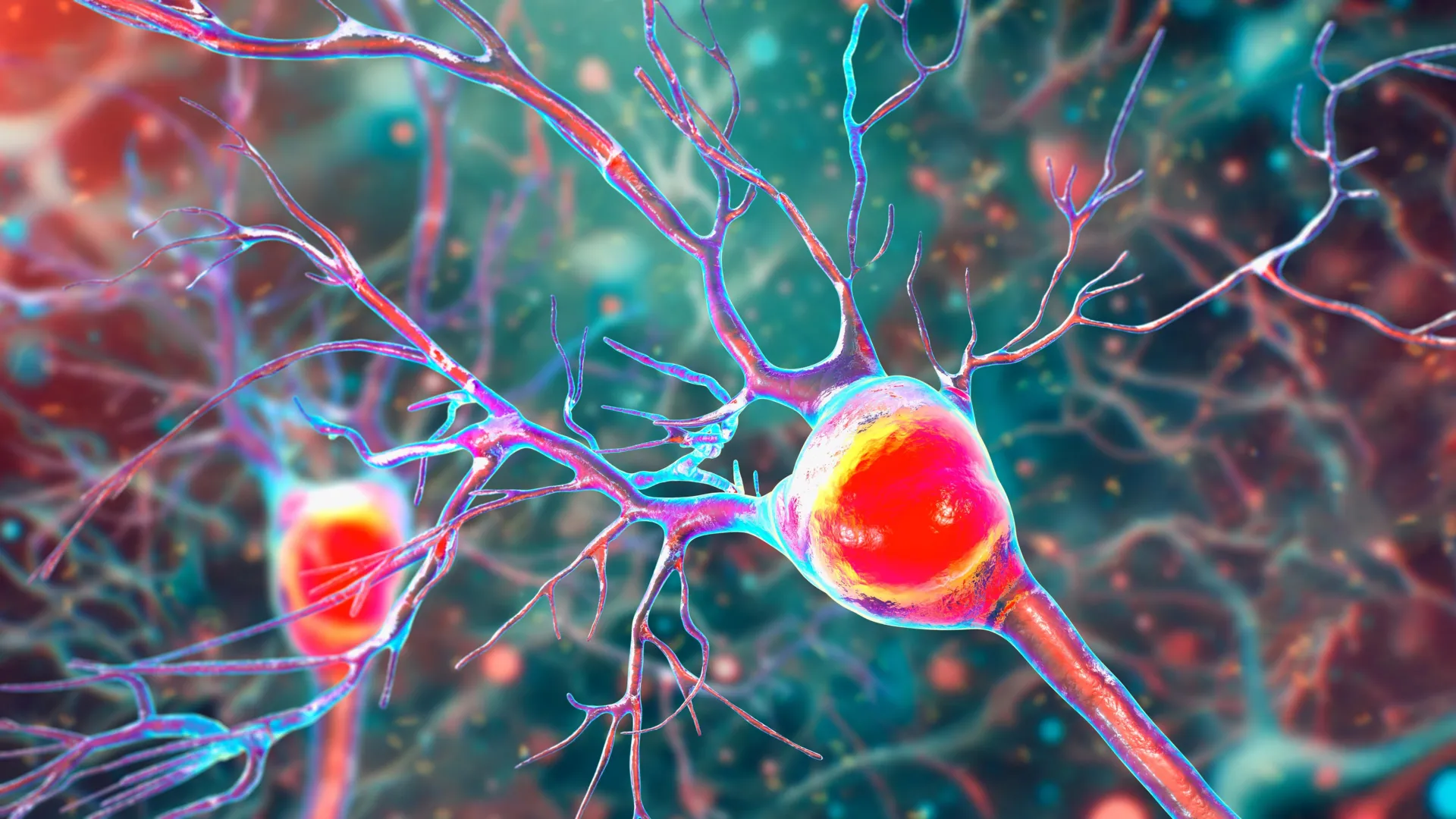
Mitochondria are often described as the "powerhouses" of the cell because they produce adenosine triphosphate (ATP), the primary energy currency of biological life. In the cerebellum, Purkinje cells are particularly vulnerable to energy shortages because they are among the largest and most metabolically active neurons in the human brain. They require a constant, high-volume supply of ATP to maintain their complex branching structures and to fire the signals that allow a person to walk, talk, or maintain balance.
The research team, led by Seema Tiwari-Woodruff, a professor of biomedical sciences at the UC Riverside School of Medicine, and graduate student Kelley Atkinson, discovered that inflammation and demyelination in the cerebellum directly disrupt mitochondrial function. Specifically, they observed a significant reduction in the mitochondrial protein COXIV (Cytochrome c oxidase subunit 4). This protein is essential for the electron transport chain, the process by which mitochondria generate energy. When COXIV levels drop, the energy supply to the Purkinje cell fails, eventually leading to the death of the neuron.
Evidence from Human Tissue and Experimental Models
To validate their findings, the UCR researchers employed a dual-track methodology, analyzing both human postmortem tissue and animal models. The human samples were obtained from individuals who had been diagnosed with secondary progressive multiple sclerosis, a stage of the disease where disability increases steadily regardless of relapses. These samples, provided by the National Institutes of Health’s NeuroBioBank and the Cleveland Clinic, were compared against cerebellar tissue from healthy donors.
The analysis of the MS-affected tissue revealed a stark reality: the Purkinje cells in these patients were not only fewer in number but also structurally compromised. The surviving neurons exhibited "dendritic pruning," a process where the branches used to receive signals from other neurons wither away. Furthermore, these cells showed a clear lack of myelin and a marked deficiency in mitochondrial markers.
To observe the chronology of this damage, the team utilized the experimental autoimmune encephalomyelitis (EAE) mouse model. EAE is a widely accepted surrogate for MS research because it mimics the T-cell-mediated autoimmune attack on the central nervous system. By tracking the progression of EAE in mice, the researchers were able to confirm that mitochondrial dysfunction begins early in the disease process. While the actual death of the Purkinje cells occurred in the later, more severe stages of the condition, the metabolic failure and demyelination were present much sooner. This suggests a "window of opportunity" for treatment before the damage becomes irreversible.
Why Purkinje Cells are Central to MS Disability
The cerebellum operates as the brain’s "quality control" center for movement. When you reach for a cup of coffee or step over a curb, the cerebellum integrates sensory input to ensure the movement is smooth and accurate. The Purkinje cells are the only neurons that send information out of the cerebellar cortex, making them the ultimate arbiters of motor coordination.
"Inside the cerebellum are special cells called Purkinje neurons," Tiwari-Woodruff explained. "These large, highly active cells help coordinate smooth, precise movements—like dancing, throwing a ball, or even just walking. They’re essential for balance and fine motor skills."
As MS progresses and these cells die off, patients develop ataxia. Ataxia is a neurological sign consisting of a lack of voluntary coordination of muscle movements that can include gait abnormality, speech changes, and abnormalities in eye movements. Because the brain has limited capacity to regenerate these complex neurons once they are lost, the resulting disability is often permanent. The UCR study’s focus on the energy failure of these cells highlights why current anti-inflammatory drugs, which reduce the number of MS relapses, often fail to stop the long-term progression of physical disability.
Broader Implications for Treatment and Research
The discovery that mitochondrial impairment contributes directly to cerebellar damage shifts the focus of MS research toward neuroprotection. Historically, MS treatments have focused on the immune system—specifically, preventing immune cells from entering the brain and attacking myelin. While these "disease-modifying therapies" (DMTs) have been successful in reducing the frequency of flare-ups, they have been less effective in the progressive forms of the disease where the primary issue is the steady loss of neurons.
The UCR findings suggest that a multi-pronged approach may be necessary. "Targeting mitochondrial health may represent a promising strategy to slow or prevent neurological decline and improve quality of life for people living with MS," Tiwari-Woodruff noted. Potential future treatments could involve "mitochondrial cocktails" or drugs designed to boost the expression of proteins like COXIV, thereby keeping the Purkinje cells alive even if the surrounding myelin is damaged.
Furthermore, the research team is expanding its scope to look at the "supporting cast" of the brain. While Purkinje cells are the primary focus of this study, they do not exist in a vacuum. They rely on oligodendrocytes to provide myelin and astrocytes to provide structural and metabolic support. Ongoing projects at UCR are investigating whether mitochondrial damage in these supporting cells also contributes to the overall environment of decay in the MS cerebellum.
A Chronology of Neurodegeneration in MS
Based on the data provided by the UCR study, a clearer timeline of cerebellar decline in MS patients is emerging:
- Initial Inflammation: The immune system targets the cerebellum, leading to an influx of inflammatory cytokines and immune cells.
- Early Demyelination: The protective myelin sheath around Purkinje cell axons is stripped away, disrupting signal conduction.
- Mitochondrial Stress: The loss of myelin and the presence of inflammatory markers lead to a decrease in COXIV protein levels.
- Metabolic Failure: The Purkinje cells can no longer produce enough ATP to maintain their complex structure, leading to the loss of dendritic branches.
- Ataxia Onset: As the neurons struggle to function, the patient begins to experience balance and coordination issues.
- Neuronal Death (Apoptosis): In the chronic and progressive stages, the energy-starved Purkinje cells die, leading to permanent loss of motor function.
The Vital Role of Sustained Scientific Funding
The UCR study, which included contributions from researchers Shane Desfor, Micah Feria, Maria T. Sekyia, Marvellous Osunde, Sandhya Sriram, Saima Noori, Wendy Rincón, and Britany Bello, was funded by the National Multiple Sclerosis Society. However, Tiwari-Woodruff used the publication of these findings to advocate for the continued support of basic and clinical science.
"Cutting funding to science only slows progress when we need it most," she stated. "Public support for research matters now more than ever."
The economic burden of multiple sclerosis in the United States alone is estimated at over $85 billion annually, including direct medical costs and indirect costs such as lost productivity. Research that leads to even a marginal delay in the progression of the disease could save billions in long-term care costs and, more importantly, preserve the independence and quality of life for millions of individuals.
Conclusion and Future Outlook
The study titled "Decreased mitochondrial activity in the demyelinating cerebellum of progressive multiple sclerosis and chronic EAE contributes to Purkinje cell loss" marks a significant step forward in the field of neuroimmunology. By identifying a specific protein deficiency (COXIV) and a specific cellular failure (mitochondrial ATP production) in the cerebellum, the research provides a concrete target for the next generation of MS drugs.
The transition from understanding MS as a disease of the immune system to a disease of cellular metabolism represents a paradigm shift. If scientists can find ways to protect the "powerhouses" of the brain, they may finally be able to offer patients more than just a reduction in relapses—they may be able to offer a way to keep walking, dancing, and moving, even in the face of a chronic diagnosis. The research team at UC Riverside continues to work toward this goal, seeking to translate these bench-top discoveries into bedside therapies that address the most debilitating aspects of multiple sclerosis.