
The study, primarily conducted in murine models and validated with human tissue samples, marks a significant milestone in neuro-oncology. By pinpointing the role of the brain’s innate immune system, the research team has not only validated the lived experience of thousands of patients but has also identified specific therapeutic targets to reverse the damage. For the first time, scientists have demonstrated that immunotherapy alone, independent of chemotherapy or radiation, is sufficient to cause lasting neurobiological changes that impair memory and executive function.
The Rise of CAR-T Therapy and the Emergence of Cognitive Side Effects
CAR-T cell therapy represents one of the most sophisticated forms of personalized medicine. The process involves harvesting a patient’s own T cells—the "soldiers" of the immune system—and genetically engineering them in a laboratory to express a specific receptor. This receptor allows the T cells to recognize and bind to a protein found on the surface of cancer cells. Once these "super-charged" cells are infused back into the patient, they proliferate and launch a targeted attack on the malignancy.
Since the U.S. Food and Drug Administration (FDA) first approved CAR-T therapy for acute lymphoblastic leukemia in 2017, its applications have expanded rapidly. It is now a standard of care for several types of non-Hodgkin lymphoma and multiple myeloma, and dozens of clinical trials are currently investigating its efficacy against solid tumors, including glioblastomas and pediatric brain stem tumors.
Despite its success in achieving long-term remission, the therapy is known for its intense systemic side effects. The most common is cytokine release syndrome (CRS), a systemic inflammatory response that can cause high fevers and organ dysfunction. Another is Immune Effector Cell-Associated Neurotoxicity Syndrome (ICANS), which can cause acute confusion, tremors, or seizures. While ICANS is typically temporary and managed in an intensive care setting, the "brain fog" described by patients is more subtle, chronic, and has historically been harder to quantify.
Uncovering a Unifying Principle of Brain Fog
The Stanford study, led by senior author Michelle Monje, MD, PhD, the Milan Gambhir Professor in Pediatric Neuro-Oncology, sought to determine if this cognitive impairment was a byproduct of the cancer itself, previous treatments, or the CAR-T cells themselves. Dr. Monje, a pioneer in the study of "chemo-brain," hypothesized that various insults to the immune system might converge on a single pathway in the brain.
To test this, the research team utilized mouse models with tumors located in diverse regions: the brain, the blood, the skin, and the bone. This variety allowed the scientists to observe whether the location of the cancer influenced the severity of cognitive decline. After administering CAR-T therapy, the researchers conducted a battery of cognitive assessments, including novel object recognition tests and maze navigation, to measure memory and spatial learning.
The results were consistent across almost all groups. Mice with brain, blood, and skin cancers all exhibited mild but measurable cognitive impairment following CAR-T treatment. The only exception was a group of mice with bone cancer that elicited a very low inflammatory response. This finding was pivotal; it suggested that the cognitive decline was not caused by the CAR-T cells directly attacking brain tissue, but rather by the systemic inflammatory "echo" produced by the immune system’s activation.
"This is the first study to demonstrate that immunotherapy on its own is sufficient to cause lasting cognitive symptoms," Dr. Monje stated. "We found the exact same pathophysiology we’ve seen in brain fog syndromes that occur after chemotherapy, radiation, and mild respiratory COVID-19 or influenza."
The Cellular Mechanism: Microglia and Myelin Damage
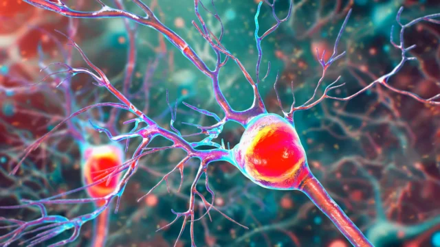
The study identified the brain’s resident immune cells, known as microglia, as the primary culprits. Under normal conditions, microglia act as the brain’s "gardeners," clearing debris and maintaining neuronal health. However, when the body undergoes a massive immune event—such as the infusion of CAR-T cells—the microglia become chronically activated, or "annoyed."
These activated microglia begin to secrete high levels of inflammatory molecules called cytokines and chemokines. These molecules create a hostile environment for other essential brain cells, specifically oligodendrocytes. Oligodendrocytes are responsible for producing myelin, the fatty insulating sheath that wraps around nerve fibers (axons). Myelin is critical for the rapid transmission of electrical signals across the brain; without it, communication between neurons slows down significantly.
The Stanford team found that the inflammatory signals from the microglia disrupted the maturation and function of oligodendrocytes, leading to a reduction in myelin density. This degradation of the brain’s "white matter" translates directly into the symptoms of brain fog: slower processing speeds, difficulty multitasking, and memory lapses.
To confirm these findings in humans, the researchers analyzed postmortem brain tissue from patients who had participated in a clinical trial for CAR-T therapy targeting diffuse intrinsic pontine glioma (DIPG), a lethal brain stem tumor. The analysis revealed the same patterns of microglial activation and oligodendrocyte dysregulation observed in the mice, providing a high degree of confidence that the mouse model accurately reflects human biology.
Strategies for Reversal and Recovery
Perhaps the most promising aspect of the study is the identification of two distinct strategies to reverse the cognitive damage. Because the impairment is driven by cellular signaling rather than the permanent death of neurons, the researchers believed the brain could be "reset."
The first strategy involved the transient depletion of microglia. By administering a compound that temporarily cleared microglia from the brains of the mice for two weeks, the researchers allowed the neural environment to stabilize. When the microglia eventually repopulated the brain, they returned in a resting, non-inflammatory state. Following this "reset," the mice performed normally on cognitive tests, and their myelin levels recovered.
The second strategy focused on blocking the specific signals sent by the "annoyed" microglia. The researchers identified a specific chemokine receptor that mediates the damage to oligodendrocytes. By administering a medication that enters the brain and interferes with these signals, they were able to rescue cognitive function without needing to deplete the immune cells themselves.
"That alone rescued cognition," Monje said, noting that these compounds are similar to drugs already in clinical development for other conditions. This similarity could significantly shorten the timeline for bringing a "brain fog" treatment to human patients.
Implications for Pediatric Oncology and Quality of Life
The study’s findings are particularly resonant for the field of pediatric oncology. As CAR-T therapy becomes a more common treatment for childhood leukemias and is tested for pediatric brain tumors, the long-term impact on the developing brain is a paramount concern.
"We’re deeply interested in how cancer therapies affect cognition because it affects patients’ quality of life," Monje emphasized. "And this is especially important for kids because their brains are still developing." For a child, a "mild" cognitive impairment can have compounding effects on learning, social development, and future independence. By understanding the mechanism of injury, clinicians can now look toward a future where the cure for cancer does not come at the cost of a child’s cognitive potential.
The research also has broader implications for "long COVID" and other post-viral syndromes. By establishing a "unifying principle" for brain fog, the study suggests that a single class of neuro-protective drugs could potentially treat cognitive impairment resulting from a wide array of triggers, from chemotherapy to severe viral infections.
Chronology of Research and Future Directions
The journey to this discovery began in 2017 with the first FDA approvals of CAR-T cell therapy. As the number of survivors grew, so did the anecdotal evidence of cognitive side effects. Dr. Monje’s lab, which had previously defined the mechanisms of "chemo-brain" in 2018 and "COVID-brain" in 2022, began applying those frameworks to immunotherapy.
The current study, led by Anna Geraghty, PhD, and Lehi Acosta-Alvarez, represents several years of intensive cross-disciplinary work involving researchers from Stanford, New York University, and Washington University in St. Louis. The project was supported by a diverse coalition of funders, including the National Institutes of Health, the Howard Hughes Medical Institute, and numerous pediatric cancer foundations such as Alex’s Lemonade Stand and the McKenna Claire Foundation.
Moving forward, the research team is focused on translating these mouse-model successes into human clinical trials. The goal is to develop a "co-therapy"—a medication taken alongside or after CAR-T treatment—that prevents or reverses microglial activation. If successful, this would allow patients to benefit from the life-saving power of immunotherapy while maintaining their mental clarity and quality of life.
The identification of a specific molecular target marks a shift in how the medical community views cognitive side effects. No longer dismissed as a vague or unavoidable consequence of "fighting for one’s life," brain fog is now a recognized, measurable, and potentially treatable neurological condition. As Monje concluded, "This study is so exciting because not only have we identified the cells central to this pathophysiology, we’ve found a molecular target we can investigate to treat it."