The identification of this benchmark comes at a critical time in neurodegenerative research, as the medical community shifts toward precision medicine and the development of therapies that target specific proteins in the brain. By establishing a quantifiable threshold for tau protein—a primary hallmark of Alzheimer’s—clinicians and researchers can more accurately distinguish between normal age-related changes and the early pathological stages of dementia.
The Biological Markers of Alzheimer’s: Amyloid and Tau
To understand the significance of the USC study, it is essential to contextualize the two primary proteins associated with Alzheimer’s pathology: amyloid-beta and tau. For decades, the "amyloid cascade hypothesis" dominated the field, suggesting that the buildup of amyloid plaques was the initial and primary driver of the disease. However, while amyloid often appears years before symptoms manifest, it has a relatively weak correlation with actual cognitive decline.
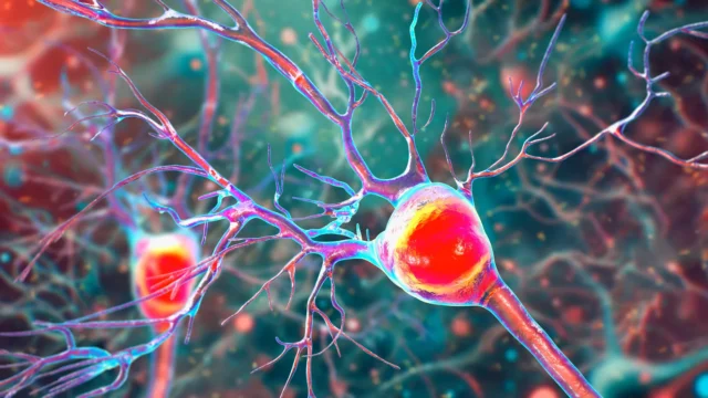
In contrast, tau protein forms "neurofibrillary tangles" inside neurons. These tangles disrupt the transport of nutrients and essential molecules, eventually leading to cell death. Crucially, the spread of tau throughout the brain’s cortex mirrors the progression of symptoms, such as memory loss and impaired judgment, much more closely than amyloid does. Because tau is the "executioner" of neurons, being able to visualize and quantify its presence is vital for diagnosing the disease and predicting its trajectory.
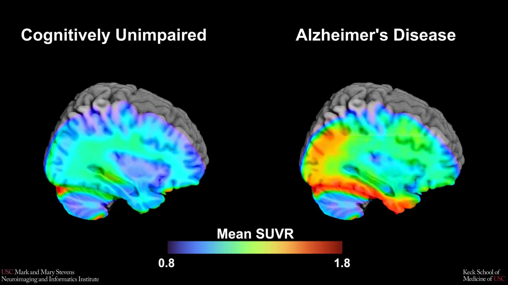
The USC research team utilized an advanced imaging technology known as tau Positron Emission Tomography (PET) to observe these proteins in living patients. This process involves injecting a specialized radioactive tracer—in this case, 18F-PI-2620—which binds to tau tangles and glows on a scan, allowing scientists to map the density and location of the protein.
Methodology and the HABS-HD Framework
The study analyzed data from over 675 older adults participating in the HABS-HD program. The Health and Aging Brain Study-Health Disparities is recognized as the most comprehensive study of Alzheimer’s disease in diverse communities. Historically, clinical trials and neuroimaging benchmarks have been developed using cohorts that were overwhelmingly non-Hispanic White, leading to diagnostic tools that may not be equally effective for all ethnic and racial groups.
The participants were categorized into two primary groups: those who were cognitively impaired and those who exhibited normal cognitive function for their age, based on standardized neuropsychological testing. The research team focused their analysis on the medial temporal lobe, a region deep within the brain that includes the hippocampus and is responsible for memory formation. This area is typically the first to show tau accumulation in the early stages of Alzheimer’s.
By comparing the PET scans of the impaired group against the control group, the researchers sought to find a "cut-point"—a mathematical threshold where the amount of tau protein becomes clinically significant.
Key Findings: Successes and Disparities
The research team successfully identified a tau cut-point in the medial temporal lobe that strongly indicated cognitive impairment. However, the data revealed complex nuances regarding how this benchmark applies across different demographics.
The study found that the tau cut-point was highly effective in distinguishing cognitive impairment in Hispanic and non-Hispanic White participants, but only when another factor was present: amyloid. In these groups, the presence of both high tau (above the cut-point) and high amyloid provided a clear biological signature of Alzheimer’s-related decline.
"Our tau cut-point was able to distinguish whether study participants had cognitive impairment—but only when another abnormal protein, amyloid, was also present in those with cognitive impairment, and only in Hispanic and non-Hispanic White participants," explained senior author Meredith N. Braskie, PhD, an assistant professor of neurology at the Keck School of Medicine.
The results took a different turn when analyzing non-Hispanic Black participants. In this cohort, the established tau cut-point did not reliably correlate with cognitive impairment. This finding is significant because it suggests that the biological drivers of dementia may vary across different racial and ethnic groups.
"In non-Hispanic Black participants, the tau cut-point did not perform as expected," Dr. Braskie noted. "This suggests that other pathologies or conditions may be driving cognitive decline in this group. Our study is an important step toward better understanding how tau relates to cognition in diverse populations."
Analyzing the Disparities: Beyond Tau and Amyloid
The failure of the tau benchmark to predict impairment in Black participants highlights a growing area of concern in neurology: the "mixed pathology" of dementia. Researchers suspect that in Black populations, cognitive decline may be more frequently influenced by vascular factors—such as hypertension, diabetes, and small vessel disease—or social determinants of health, rather than the traditional amyloid-tau progression seen in other groups.
Lead author Victoria R. Tennant, a PhD candidate in USC’s Neuroscience Graduate Program, emphasized that the findings underscore the danger of a "one-size-fits-all" approach to Alzheimer’s diagnosis. "The limited reliability of tau as an indicator of cognitive impairment in non-Hispanic Black participants highlights the need for more diverse populations in research and for future studies to examine both biological and social determinants of Alzheimer’s disease," Tennant said.
This discrepancy has profound implications for clinical trials. Many of the newest Alzheimer’s treatments, such as monoclonal antibodies, are designed to clear amyloid or tau from the brain. If a patient’s cognitive decline is being driven by vascular issues rather than tau tangles, these expensive and potentially side-effect-heavy treatments may be ineffective.
The Role of the 18F-PI-2620 Tracer
A technical highlight of the study was the use of the second-generation imaging tracer 18F-PI-2620. Earlier tau tracers often suffered from "off-target binding," where the tracer would stick to other proteins or structures in the brain, creating "noise" that made it difficult to get an accurate reading of tau levels.
The 18F-PI-2620 tracer is engineered for higher specificity, meaning it binds more cleanly to the tau tangles associated with Alzheimer’s. By using this more precise tool, the USC team was able to define a more reliable threshold for what constitutes "tau positivity." This technical advancement is a necessary step for moving tau PET scans from the realm of pure research into everyday clinical practice.
Chronology of Progress at Stevens INI
The Mark and Mary Stevens Neuroimaging and Informatics Institute has been at the forefront of Alzheimer’s research for years. This latest study is part of a chronological series of breakthroughs facilitated by the HABS-HD initiative.
- Early 2010s: Recognition of the "diversity gap" in Alzheimer’s research leads to the conceptualization of HABS-HD.
- 2016-2018: The University of North Texas Health Science Center, in collaboration with USC and other partners, begins recruiting a diverse cohort of thousands of participants.
- 2020-2022: Initial findings from HABS-HD identify that Hispanic populations may develop Alzheimer’s symptoms at an earlier age, often linked to metabolic factors.
- 2023-2024: The focus shifts to neuroimaging biomarkers, leading to the current study on tau cut-points and the realization that tau’s role varies by ethnicity.
Arthur W. Toga, PhD, director of the Stevens INI, noted that these findings are part of a larger mission to provide personalized care. "This type of imaging is critical for understanding who is at risk and how the disease develops," Toga said. "HABS-HD has already produced key findings related to ethnic variations in AD biomarkers, the influences of social determinants on cognitive health, and vascular contributions to dementia."
Broader Impact and Future Implications
The establishment of tau cut-points is more than an academic exercise; it is a foundational requirement for the next generation of Alzheimer’s care. As the FDA continues to approve new disease-modifying therapies, the ability to identify the right patient for the right drug is paramount.
For the Hispanic and non-Hispanic White communities, this study provides a concrete metric that clinicians can eventually use to say, "Your tau levels have crossed the threshold; it is time to consider intervention." For the Black community, the study serves as a call to action for researchers to look deeper into the specific biological and environmental factors that contribute to brain aging.
Furthermore, the study emphasizes that Alzheimer’s is a staged disease. By the time memory loss is obvious, the brain has already undergone significant damage. Tools like the tau PET cut-point allow for "pre-symptomatic" detection, opening a window for preventative strategies and early-stage clinical trial enrollment.
The research was a massive collaborative effort, involving experts from the Alzheimer’s Therapeutic Research Institute at USC, the Waisman Center at the University of Wisconsin-Madison, Washington University School of Medicine in St. Louis, and the University of California, San Francisco. This multi-disciplinary approach ensures that the findings are robust and vetted across different scientific perspectives.
Conclusion
The Keck School of Medicine’s identification of a tau PET benchmark represents a significant leap forward in the quest to demystify Alzheimer’s disease. While it provides a powerful new tool for identifying risk in Hispanic and White populations, it also highlights the urgent need for a more inclusive understanding of cognitive decline in Black populations. As the HABS-HD project continues to expand, the data gathered will likely lead to a more nuanced, equitable, and effective framework for treating one of the most challenging diseases of the 21st century.
This research was supported by several grants from the National Institute on Aging of the National Institutes of Health, including R01AG054073 and U19AG078109, as well as support from the NIH Office of the Director. These investments reflect the national priority placed on solving the Alzheimer’s crisis through diverse and technologically advanced scientific inquiry.