
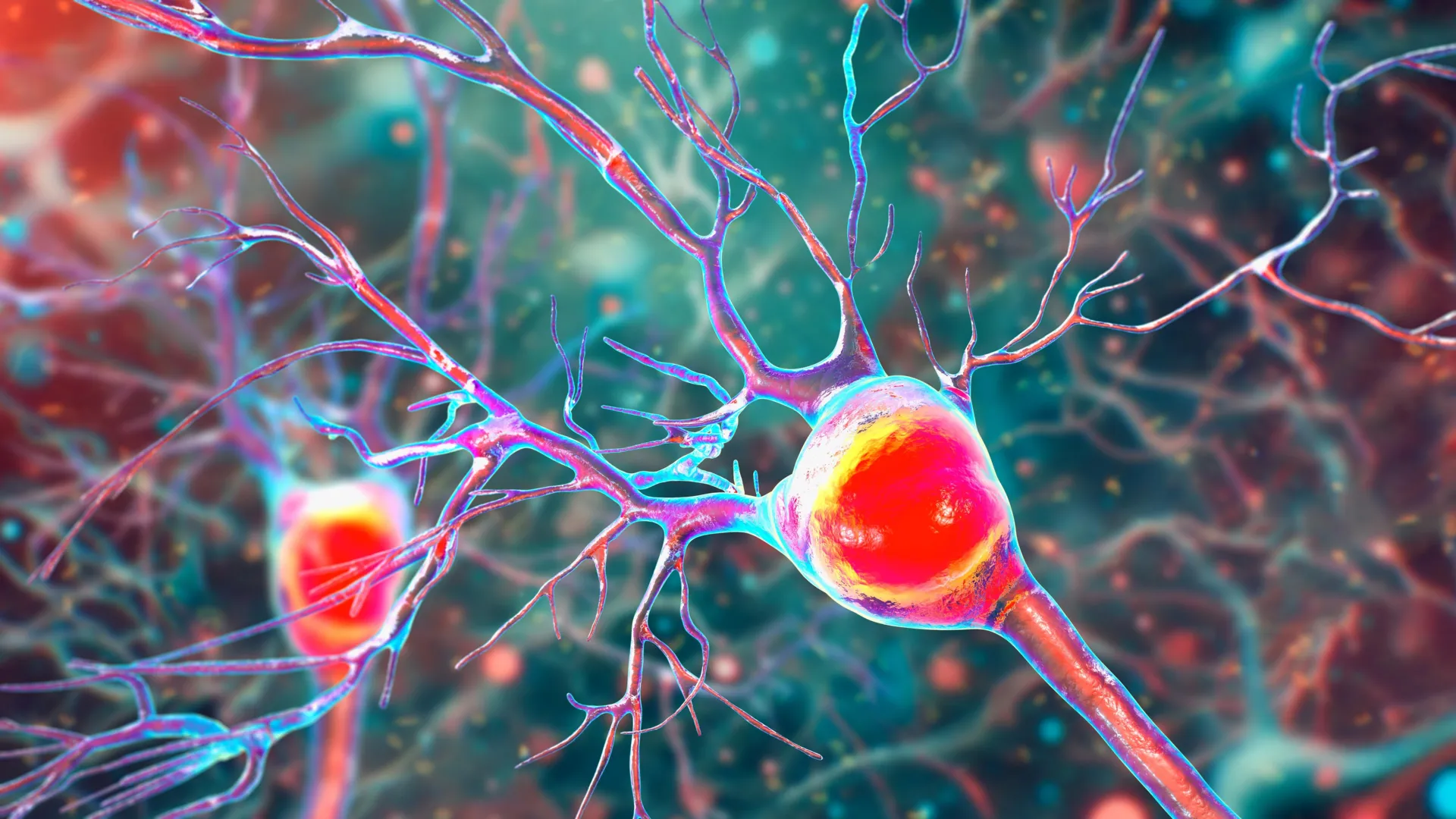
A groundbreaking study led by researchers at the University of California, Riverside (UCR) has provided a new understanding of the cellular mechanisms driving this neurological decline. Published in the Proceedings of the National Academy of Sciences (PNAS), the research identifies a critical link between malfunctioning mitochondria and the death of Purkinje cells—large, specialized neurons that are essential for cerebellar function. The study suggests that the progressive breakdown of these cells is not merely a byproduct of inflammation but is driven by a localized energy crisis within the neurons themselves.
The Biological Architecture of Cerebellar Decline
The central nervous system relies on a delicate balance of electrical signaling and metabolic support. In a healthy brain, nerve fibers are insulated by the myelin sheath, a lipid-rich layer that facilitates the rapid transmission of electrical impulses. Multiple sclerosis is fundamentally characterized by demyelination, an autoimmune process where the body’s immune system mistakenly attacks and destroys this protective coating. Without myelin, the efficiency of signal conduction drops significantly, leading to the sensory and motor deficits characteristic of the disease.
However, demyelination is only one part of the equation. To maintain their high level of activity, neurons require a constant and robust supply of energy. This energy is produced by mitochondria, the "powerhouses" of the cell, through a process known as oxidative phosphorylation. The UCR research team, led by Seema Tiwari-Woodruff, a professor of biomedical sciences at the UC Riverside School of Medicine, proposes that the intersection of inflammation and demyelination creates a toxic environment that directly impairs mitochondrial health.
"Our study, conducted by my graduate student Kelley Atkinson, proposes that inflammation and demyelination in the cerebellum disrupt mitochondrial function, contributing to nerve damage and Purkinje cell loss," stated Professor Tiwari-Woodruff. The team specifically observed a marked reduction in the mitochondrial protein COXIV within demyelinated Purkinje cells. COXIV is a vital component of the electron transport chain, and its absence indicates a fundamental failure in the cell’s ability to produce adenosine triphosphate (ATP), the primary energy currency of biological life.
The Vital Role of Purkinje Neurons in Motor Coordination
To understand the gravity of these findings, it is necessary to consider the unique role of Purkinje cells. These neurons are among the largest in the human brain and possess an incredibly complex "dendritic tree"—a vast network of branches that receive and process thousands of inputs from other parts of the nervous system. Every physical action, from the rhythmic motion of walking to the precise finger movements required for typing or playing a musical instrument, is mediated by the inhibitory signals sent by Purkinje cells to the deep cerebellar nuclei.
When Purkinje cells are compromised, the brain loses its ability to refine motor commands. This leads to a condition known as ataxia, characterized by jerky, uncoordinated movements and a lack of balance. "Inside the cerebellum are special cells called Purkinje neurons," Tiwari-Woodruff explained. "These large, highly active cells help coordinate smooth, precise movements—like dancing, throwing a ball, or even just walking. They’re essential for balance and fine motor skills."
The UCR study highlights that in the context of MS, these cells undergo a tragic transformation. Analyzing brain tissue from deceased MS patients, the researchers found that surviving Purkinje neurons exhibited fewer branches and significant myelin loss. Most importantly, their mitochondria were failing, effectively starving the cells of the energy required to maintain their structure and function. This metabolic exhaustion eventually triggers programmed cell death, leading to a permanent reduction in the brain’s motor-processing capacity.
Chronology of Disease Progression: Insights from Mouse Models
To track the specific timeline of how mitochondrial failure leads to cell death, the research team utilized the experimental autoimmune encephalomyelitis (EAE) mouse model. EAE is the gold-standard laboratory model for studying MS, as it mimics the inflammatory and demyelinating processes seen in human patients.
By observing the progression of the disease in these models, the researchers were able to establish a clear chronology of degeneration:
- Early Phase: Rapid demyelination occurs within the cerebellar white matter. During this stage, the mice begin to show the first signs of motor instability, though the neurons remain physically present.
- Intermediate Phase: As demyelination persists, the mitochondrial health of the Purkinje cells begins to decline. The loss of COXIV protein becomes evident, and the energy output of the cells drops.
- Late Phase: The metabolic crisis reaches a breaking point. Deprived of energy and stripped of their protective myelin, the Purkinje cells begin to die off in large numbers. This stage correlates with the most severe clinical symptoms, including total loss of coordination.
"We saw that the myelin breaks down early in the disease," Tiwari-Woodruff noted. "These problems—less energy, loss of myelin, and damaged neurons—start early, but the actual death of the brain cells tends to happen later, as the disease becomes more severe. The loss of energy in brain cells seems to be a key part of what causes damage in MS."
This timeline is significant because it identifies a "window of opportunity" for medical intervention. If mitochondrial function can be supported or restored during the early or intermediate phases, it may be possible to prevent the irreversible loss of neurons that occurs in the later stages of the disease.
Comparative Analysis and Supporting Data
The strength of the UCR study lies in its dual approach, combining laboratory models with the analysis of human tissue. The researchers examined postmortem cerebellar tissue from individuals who had been diagnosed with secondary progressive MS, a stage of the disease where disability increases steadily over time. These samples were compared against tissue from healthy donors, provided by the National Institutes of Health’s NeuroBioBank and the Cleveland Clinic.
The data revealed a stark contrast:
- Neuron Density: MS patients showed a significantly lower density of Purkinje cells compared to healthy controls, particularly in areas where demyelination was most severe.
- Mitochondrial Markers: The expression of mitochondrial proteins was markedly lower in the MS samples, confirming that the "energy failure" observed in mice is a direct reflection of the human disease state.
- Structural Integrity: The dendritic trees of the remaining Purkinje cells in MS patients were stunted and less complex, limiting their ability to communicate with other neurons.
These findings reinforce the theory that MS is not just a disease of the immune system, but also a disease of metabolic failure. While current MS treatments largely focus on suppressing the immune system to prevent new inflammatory attacks, they often do little to address the underlying neurodegeneration that continues in the absence of active inflammation.
Future Therapeutic Horizons and Broader Implications
The identification of mitochondrial failure as a primary driver of Purkinje cell loss opens new avenues for therapeutic development. Future treatments may focus on "neuroprotection"—strategies designed to keep neurons alive and functional despite the presence of the disease.
Professor Tiwari-Woodruff and her team are already looking toward the next phase of research. They are investigating whether mitochondrial damage extends to other critical cell types in the cerebellum, such as:
- Oligodendrocytes: The cells responsible for producing myelin. If their mitochondria fail, the brain loses its ability to repair the damage caused by MS.
- Astrocytes: Support cells that provide nutrients to neurons and maintain the blood-brain barrier.
"Targeting mitochondrial health may represent a promising strategy to slow or prevent neurological decline and improve quality of life for people living with MS," said Tiwari-Woodruff. Potential interventions could include drugs that boost mitochondrial biogenesis, antioxidants that protect the energy-producing structures from oxidative stress, or metabolic supplements that provide alternative energy sources to struggling neurons.
The implications of this research extend beyond MS. Mitochondrial dysfunction is a common thread in many neurodegenerative conditions, including Parkinson’s disease and Alzheimer’s disease. By solving the mystery of Purkinje cell loss in the cerebellum, researchers may uncover fundamental truths about how the brain responds to chronic stress and energy depletion.
The Necessity of Sustained Research Support
The study was a collaborative effort involving several researchers, including Shane Desfor, Micah Feria, Maria T. Sekyia, Marvellous Osunde, Sandhya Sriram, Saima Noori, Wendy Rincán, and Britany Belloa. The work was made possible through funding from the National Multiple Sclerosis Society, highlighting the vital role of non-profit and public support in advancing medical science.
In her concluding remarks, Professor Tiwari-Woodruff emphasized the importance of continued investment in the scientific community. She warned that the pace of discovery is directly tied to the availability of resources. "Cutting funding to science only slows progress when we need it most," she said. "Public support for research matters now more than ever."
As the scientific community moves closer to understanding the complex interplay between the immune system and cellular metabolism, the hope for more effective, targeted treatments for MS grows. For the millions of people living with the daily challenges of balance and coordination issues, this research represents a critical step toward a future where the progression of the disease can be halted, and the "powerhouses" of the brain can be restored to full health.