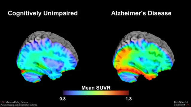
Alzheimer’s disease remains the most prevalent cause of dementia worldwide, characterized by the progressive accumulation of toxic proteins that disrupt neural communication. For decades, the medical community has sought non-invasive and cost-effective methods to identify the disease in its prodromal—or pre-symptomatic—stages. The NTU Singapore findings suggest that the brain’s "drainage pipes" offer a clear visual map of this impending decline.
The Biological Mechanism of Brain Waste Clearance
To understand the significance of the study, one must look at the glymphatic system and the perivascular spaces that facilitate the brain’s internal plumbing. These spaces are fluid-filled channels surrounding the blood vessels that penetrate the brain parenchyma. Their primary function is to act as a conduit for the clearance of metabolic waste products, most notably beta-amyloid and tau proteins.
In a healthy brain, these proteins are efficiently flushed out. However, in individuals predisposed to or in the early stages of Alzheimer’s, this clearance mechanism falters. As waste products accumulate, the perivascular spaces become dilated and "clogged," resulting in the "enlarged" appearance visible on an MRI. While these enlarged spaces have been noted in medical literature previously, their specific correlation with Alzheimer’s biomarkers—rather than just general aging or vascular issues—has remained a subject of intense scientific inquiry until now.
Addressing the Research Gap in Asian Populations
One of the most significant aspects of the NTU Singapore study is its focus on Asian demographics. Historically, the vast majority of Alzheimer’s research has been conducted on Caucasian cohorts in North America and Europe. This geographic bias has created a "data void," as genetic and lifestyle factors vary significantly across ethnic groups.
The NTU team, led by Associate Professor Nagaendran Kandiah, Director of the Dementia Research Centre (Singapore) at LKCMedicine, emphasized that dementia does not manifest uniformly across the globe. For instance, the apolipoprotein E4 (APOE4) gene is a well-known high-risk factor for Alzheimer’s. In Caucasian populations with dementia, the prevalence of this gene is approximately 50 to 60 percent. Conversely, among Singaporean dementia patients, the prevalence is less than 20 percent.
"Because of these differences, findings in one population may not directly apply to another," explained Assoc Prof Kandiah. To rectify this, the team analyzed a diverse group of nearly 1,000 Singaporeans, reflecting the nation’s multi-ethnic Chinese, Malay, and Indian composition. This inclusive approach ensures that the diagnostic markers identified are relevant to the millions of people across Asia facing a rising tide of age-related cognitive decline.
Methodology and Comparative Analysis
The research was a rigorous undertaking involving both neuroimaging and advanced blood-based biomarker analysis. The study participants were categorized into two primary groups: those with normal cognitive function and those exhibiting signs of mild cognitive impairment (MCI). MCI is often considered a transitional stage between the expected cognitive decline of normal aging and the more serious deficit of dementia.
The research team utilized 3-Tesla MRI scans to visually identify and quantify enlarged perivascular spaces. Simultaneously, they analyzed seven specific biochemical markers in the participants’ blood. These markers included various forms of beta-amyloid and tau proteins, as well as indicators of neuroaxonal damage.
The results were striking. Participants with MCI were significantly more likely to exhibit enlarged perivascular spaces compared to their cognitively healthy counterparts. Furthermore, the presence of these "clogged drains" was strongly correlated with four of the seven blood-based Alzheimer’s biomarkers. This correlation suggests that EPVS is not merely a sign of aging, but a direct reflection of the pathological processes—such as amyloid plaque buildup and tau tangles—that define Alzheimer’s disease.
Beyond White Matter: A More Sensitive Signal
For years, clinicians have relied on "white matter hyperintensities"—visible damage to the brain’s network of nerve fibers—as a primary indicator of small vessel disease and potential dementia. While the NTU study confirmed that white matter damage is indeed linked to Alzheimer’s markers, it revealed a more nuanced truth.
Among the participants with early cognitive decline, the link between Alzheimer’s-related biochemicals and enlarged perivascular spaces was found to be stronger than the link with white matter damage. This suggests that EPVS may serve as a more sensitive and earlier signal of neurodegeneration.
"Although white matter damage is more widely used in clinical practice to evaluate for dementia, as it is easily recognized on MRI scans, our results suggest that enlarged perivascular spaces may hold unique value in detecting early signs of Alzheimer’s disease," said Assoc Prof Kandiah.
Clinical Implications and Expert Reactions
The ability to use routine MRI scans for early detection carries profound implications for healthcare systems. Currently, definitive Alzheimer’s diagnosis often requires expensive Positron Emission Tomography (PET) scans or invasive lumbar punctures to check cerebrospinal fluid. By using EPVS as a marker on standard MRIs, doctors can identify high-risk patients without the need for additional, costly tests.
Justin Ong, the study’s first author and a fifth-year medical student at LKCMedicine, highlighted the importance of this timing. "Identifying Alzheimer’s sooner gives doctors more time to intervene and potentially slow the progression of symptoms such as memory loss, reduced thinking speed, and mood changes," Ong noted.
External experts have also weighed in on the study’s potential. Dr. Rachel Cheong Chin Yee, a Senior Consultant at Khoo Teck Puat Hospital’s Department of Geriatric Medicine, noted that the research clarifies the role of small blood vessel changes in the development of the disease. She emphasized that these findings could help identify high-risk individuals even before the first symptoms of memory loss appear.
Dr. Chong Yao Feng, a Consultant at the National University Hospital’s Division of Neurology, pointed out that the study challenges the traditional separation of cerebrovascular disease (blood vessel issues) and Alzheimer’s disease (protein issues). "The study’s findings are intriguing as they demonstrate that both diseases do interact in a synergistic manner," Dr. Chong said. He advised that when doctors see these enlarged spaces on a scan, they should no longer assume it is "just" a vascular issue, but rather a potential indicator of underlying Alzheimer’s pathology.
Chronology of the Research and Future Directions
The research originated as part of the Scholarly Project module within the Bachelor of Medicine and Bachelor of Surgery (MBBS) programme at LKCMedicine. This highlights the school’s commitment to integrating high-level research into its medical curriculum. Following the initial data collection and analysis, the findings have now been presented to the global scientific community, marking a milestone in Singaporean neuroscience.
The next phase of the research is already underway. The NTU team plans to conduct a longitudinal study, tracking the 1,000 participants over several years. This follow-up will allow researchers to see exactly how many individuals with enlarged perivascular spaces eventually progress to a clinical diagnosis of Alzheimer’s dementia.
If the longitudinal data confirms the predictive power of these "clogged drains," it could lead to a global shift in how MRIs are interpreted. Radiologists may soon be trained to specifically look for and grade EPVS as a standard part of cognitive assessments.
Conclusion: A New Frontier in Neurodegeneration
As the global population ages, the burden of Alzheimer’s disease is expected to triple by 2050. In Asia, where the aging transition is happening more rapidly than in the West, the need for accessible diagnostic tools is urgent. The discovery by NTU Singapore scientists provides a tangible, visual marker for a disease that has long been "invisible" until it is too late to treat effectively.
By turning a routine imaging procedure into a powerful early-warning system, this research offers hope for millions. It paves the way for a future where Alzheimer’s is managed not just at the stage of memory loss, but at the very first sign of a "clog" in the brain’s intricate drainage system. This shift from reactive treatment to proactive management could fundamentally change the trajectory of dementia care for generations to come.