
The Science of Tau and the Search for Diagnostic Precision
Alzheimer’s disease is characterized by the accumulation of two primary abnormal proteins in the brain: amyloid-beta, which forms plaques between neurons, and tau, which forms tangles inside the neurons. While amyloid has long been the primary focus of diagnostic research, recent years have seen a shift toward tau protein analysis. Tau is more closely correlated with the actual onset of cognitive decline and memory loss, making it a critical "biological clock" for the progression of the disease.
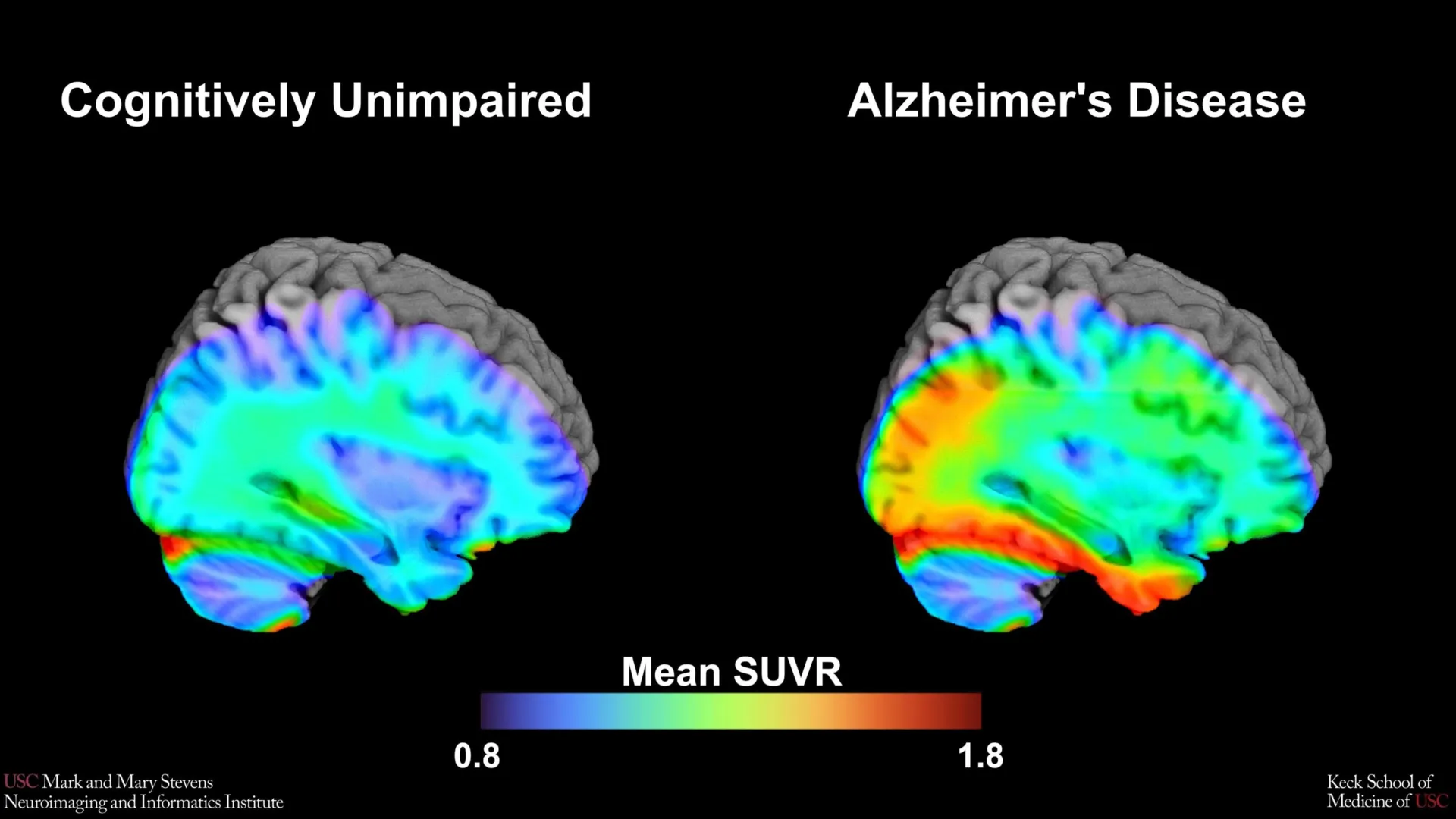
To visualize these proteins in living patients, researchers utilize Positron Emission Tomography (PET) scans. In this study, the USC team employed an advanced imaging tracer known as 18F-PI-2620. This tracer is designed to bind specifically to tau protein aggregates, allowing it to "light up" on a scan. By quantifying the amount of tracer present, researchers can determine the density of tau in specific brain regions.
The primary objective of the USC study was to establish a "tau cut-point"—a numerical threshold that distinguishes a "normal" amount of tau associated with healthy aging from a "pathological" amount indicative of Alzheimer’s disease. Using data from over 675 older adults enrolled in the HABS-HD study, the team focused on the medial temporal lobe, a region deep within the brain that serves as the epicenter for memory formation and is typically the first area affected by tau pathology.
Ethnic Disparities in Biomarker Reliability
One of the most significant contributions of this research is its focus on diversity. For decades, Alzheimer’s research has been criticized for relying on predominantly non-Hispanic White cohorts, which often fails to account for the biological and social variances found in other ethnic groups. The HABS-HD study was specifically designed to address this gap, recruiting a diverse participant pool to ensure diagnostic tools are equitable.
The USC researchers compared the tau PET scans of cognitively impaired participants with those who showed no impairment. Their findings revealed a complex landscape of diagnostic efficacy. The established tau cut-point was highly effective in identifying cognitive impairment in Hispanic and non-Hispanic White participants, but only when another abnormal protein, amyloid, was also present. This suggests that in these populations, the synergy between amyloid and tau remains a primary driver of the disease.
However, the results for non-Hispanic Black participants told a different story. In this group, the tau cut-point did not perform as a reliable indicator of cognitive impairment. This discrepancy is a major finding, suggesting that the biological drivers of cognitive decline may differ across ethnic lines. For Black participants, factors other than tau—such as vascular disease, chronic inflammation, or social determinants of health—might play a more prominent role in the development of dementia.
Analyzing the Impact of the 18F-PI-2620 Tracer
The choice of the 18F-PI-2620 tracer was pivotal to the study’s success. Unlike older generations of PET tracers, 18F-PI-2620 offers higher sensitivity and lower "off-target" binding, meaning it is less likely to stick to parts of the brain where tau is not present. This precision allowed the researchers to isolate the signal coming from the medial temporal lobe with unprecedented clarity.
"While our findings support prior research linking medial temporal lobe tau to cognitive impairment, establishing a cut-point in this region using 18F-PI-2620 marks an important step toward defining tau positivity for both research and clinical applications," said lead author Victoria R. Tennant, a PhD candidate in USC’s Neuroscience Graduate Program.
By defining what "tau positive" looks like in a diverse population, the study provides a roadmap for future clinical trials. If a drug is designed to clear tau protein from the brain, researchers must first have a reliable way to identify who actually has high levels of tau. The USC benchmark provides that gateway, ensuring that clinical trial participants are correctly categorized based on their biological profile rather than just their symptoms.
A Chronology of Progress in Alzheimer’s Diagnostics
To understand the weight of this study, it is essential to view it within the timeline of Alzheimer’s research evolution:
- Pre-2000s: Alzheimer’s could only be definitively diagnosed through an autopsy. Clinical diagnosis in living patients was based purely on behavioral observations and cognitive tests, which were often imprecise.
- Early 2000s: The advent of Amyloid PET imaging allowed researchers to see plaques in living brains for the first time. However, many people with amyloid plaques never develop dementia, leading to diagnostic confusion.
- 2010s: The development of Tau PET tracers began to bridge the gap between brain pathology and clinical symptoms. Researchers realized that tau, not amyloid, was the better predictor of when a patient would start losing their memory.
- 2020-Present: The focus has shifted toward "biomarker-based" diagnosis. The National Institute on Aging and the Alzheimer’s Association (NIA-AA) proposed a framework where AD is defined by the presence of Amyloid (A), Tau (T), and Neurodegeneration (N)—the ATN framework.
- The Current Study: The USC findings refine the "T" (Tau) component of the ATN framework by providing a specific, validated cut-point and highlighting that this "T" marker may not mean the same thing for every ethnic group.
Institutional Perspectives and Future Implications
The implications of this study extend far beyond the laboratory. Arthur W. Toga, PhD, director of the Stevens INI, emphasized the role of advanced imaging in transforming patient outcomes. "This type of imaging is critical for understanding who is at risk and how the disease develops," Toga stated. "HABS-HD has already produced key findings related to ethnic variations in AD biomarkers, the influences of social determinants on cognitive health, and vascular contributions to dementia. We hope this work will lead to more personalized care and better outcomes for all communities."
Senior author Meredith N. Braskie, PhD, assistant professor of neurology at the Keck School of Medicine, noted that the study’s failure to find a reliable tau cut-point for Black participants is not a setback, but a vital discovery. "This suggests that other pathologies or conditions may be driving cognitive decline in this group," Braskie explained. "Our study is an important step toward better understanding how tau relates to cognition in diverse populations and has important implications for future clinical trials that aim to target tau."
For clinicians, these findings suggest a move toward a more holistic diagnostic approach. If a Black patient presents with cognitive decline but does not show high levels of tau on a PET scan, the clinician might look toward vascular health, such as hypertension or small vessel disease, as the primary culprit. This prevents misdiagnosis and ensures that patients receive treatments that actually address the underlying cause of their condition.
Supporting Data and Statistical Context
The scope of the Alzheimer’s crisis underscores the urgency of this research. According to the Alzheimer’s Association:
- More than 6.7 million Americans are currently living with Alzheimer’s.
- By 2050, this number is projected to rise to nearly 13 million.
- Black Americans are approximately twice as likely to have Alzheimer’s or other dementias as older White Americans.
- Hispanics are about 1.5 times as likely to have the disease as White Americans.
Despite these statistics, minority groups have historically been underrepresented in the clinical trials used to approve new treatments. The HABS-HD initiative, and specifically this USC study, provides the data necessary to ensure that new diagnostic benchmarks are not just accurate for a subset of the population, but for the population as a whole.
The use of the 675-adult cohort provides a robust statistical foundation. By analyzing participants across the cognitive spectrum—from those with normal cognition to those with Mild Cognitive Impairment (MCI) and dementia—the researchers were able to create a gradient of tau accumulation. The finding that the medial temporal lobe tau only indicated impairment when amyloid was also present reinforces the "dual-pathology" theory of Alzheimer’s, where amyloid acts as a catalyst that allows tau to spread like wildfire across the brain.
Conclusion: The Path Toward Personalized Neurology
The USC study marks a transition from a "one-size-fits-all" diagnostic model to a more nuanced, personalized approach. By identifying the specific threshold where tau protein becomes a clinical concern, researchers have given doctors a new tool to identify Alzheimer’s in its earliest, most treatable stages.
However, the research also serves as a sobering reminder of the work yet to be done. The "biomarker gap" found in non-Hispanic Black participants highlights the necessity of investigating the social and environmental factors—such as access to healthcare, diet, and systemic stress—that may interact with biology to produce different disease profiles.
As the medical community moves closer to a future where Alzheimer’s is managed as a chronic condition rather than a terminal diagnosis, benchmarks like the tau cut-point will be essential. They represent the bridge between raw imaging data and actionable medical decisions, ensuring that every patient, regardless of their ethnic background, receives a diagnosis that is as accurate and personalized as possible. The work of the Stevens INI and the HABS-HD collaboration continues to push the boundaries of what is possible in neuroimaging, bringing the world one step closer to a future without the shadow of Alzheimer’s.