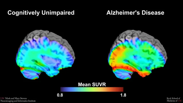
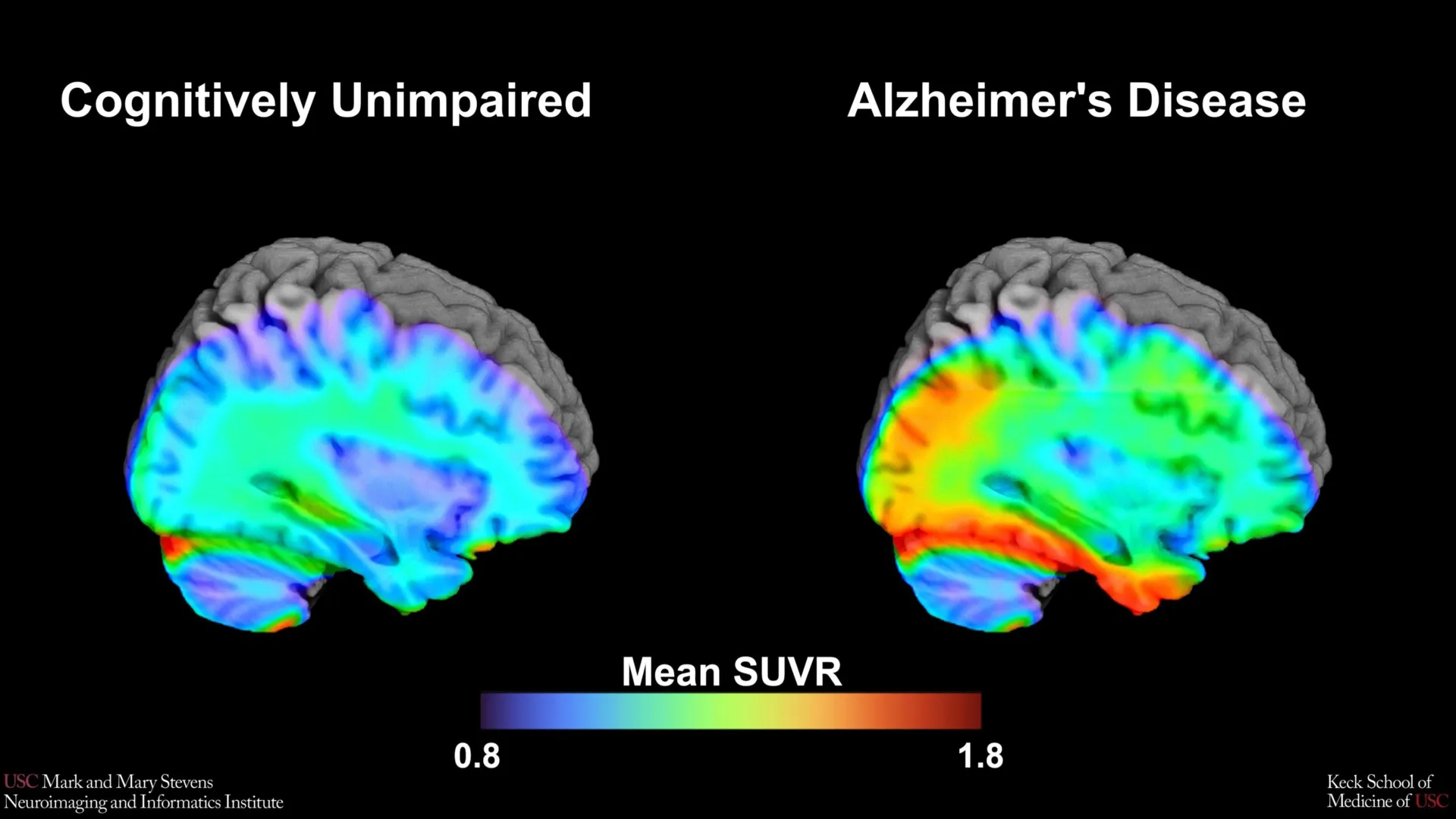
The breakthrough centers on the use of Tau Positron Emission Tomography (PET) scans, an advanced imaging modality that allows scientists to visualize the physical architecture of dementia-related proteins within a living brain. By analyzing a cohort of over 675 older adults, the USC-led team sought to define the specific threshold—or "cut-point"—at which tau protein levels transition from normal age-related changes to clinically significant markers of Alzheimer’s disease. While the study successfully identified this benchmark, it also uncovered critical discrepancies in how these biomarkers manifest across different racial and ethnic groups, highlighting a pressing need for more inclusive medical research.
The Biological Mechanics of Alzheimer’s: Amyloid vs. Tau
To understand the significance of the new tau benchmark, it is essential to distinguish between the two primary proteins associated with Alzheimer’s disease: amyloid-beta and tau. For decades, the "amyloid hypothesis" dominated the field, suggesting that the buildup of amyloid plaques was the primary driver of the disease. However, while amyloid often appears years or even decades before symptoms emerge, it is a relatively poor predictor of actual cognitive decline.
Tau protein, conversely, forms "tangles" inside neurons. Unlike amyloid, the spread of tau throughout the brain’s cortex tracks closely with the onset and progression of memory loss and functional impairment. In the "stages" of Alzheimer’s progression, amyloid is often viewed as the "trigger," while tau is the "bullet" that causes widespread neuronal death. The ability to accurately measure tau via PET imaging is therefore considered the "holy grail" of Alzheimer’s diagnostics, as it allows clinicians to see the disease’s active destructive phase.
The USC study utilized a cutting-edge radioactive tracer known as 18F-PI-2620. This tracer is designed to bind specifically to tau aggregates, allowing the PET scanner to highlight areas of accumulation. By focusing on the medial temporal lobe—a region deep within the brain that serves as the epicenter for memory formation—the researchers were able to quantify the exact amount of tau that distinguishes a "cognitively normal" brain from one experiencing the early stages of Alzheimer’s-related impairment.
Methodology and the HABS-HD Framework
The Health and Aging Brain Study-Health Disparities (HABS-HD) represents one of the most comprehensive efforts to date to study Alzheimer’s within the context of a diverse society. Historically, the vast majority of Alzheimer’s research has been conducted on non-Hispanic White populations, leaving a significant gap in the medical community’s understanding of how the disease affects Black and Hispanic communities, who are statistically at a higher risk for dementia.
For this study, the research team analyzed 675 participants, categorized by both their cognitive status (impaired vs. unimpaired) and their ethnic background. Each participant underwent a rigorous battery of cognitive tests and advanced neuroimaging. The primary goal was to establish a universal "tau cut-point"—a numerical value that would serve as a diagnostic "red line."
The results were revealing. The researchers found that the tau cut-point was highly effective at identifying cognitive impairment in Hispanic and non-Hispanic White participants, but only when another factor was present: amyloid. This "dual-biomarker" requirement suggests that in these populations, the combination of amyloid plaques and high tau levels in the medial temporal lobe is a definitive signature of Alzheimer’s disease.
Addressing the Racial Gap in Biomarker Efficacy
Perhaps the most significant finding of the study was the lack of reliability of the tau cut-point in non-Hispanic Black participants. In this group, the established threshold for tau protein did not correlate with cognitive impairment in the same way it did for White and Hispanic participants. This discrepancy suggests that the biological drivers of memory loss may differ across populations.
"In non-Hispanic Black participants, the tau cut-point did not perform as expected," noted senior author Meredith N. Braskie, PhD, an assistant professor of neurology at USC. "This suggests that other pathologies or conditions may be driving cognitive decline in this group. Our study is an important step toward better understanding how tau relates to cognition in diverse populations and has important implications for future clinical trials that aim to target tau."
Experts suggest that in Black populations, cognitive decline may be more heavily influenced by vascular contributions—such as hypertension or small vessel disease—or other proteinopathies like TDP-43, rather than the traditional amyloid-tau pathway. This finding challenges the "one-size-fits-all" approach to Alzheimer’s diagnostics and underscores the necessity of personalized medicine that accounts for a patient’s specific biological and social background.
A Chronology of Alzheimer’s Imaging Advancements
The development of the tau cut-point is the latest milestone in a timeline of rapid diagnostic evolution:
- Pre-2000s: Alzheimer’s could only be definitively diagnosed post-mortem through an autopsy. Clinical diagnosis in living patients was based entirely on behavioral symptoms.
- 2004: The introduction of "Pittsburgh Compound B" (PiB), the first PET tracer capable of imaging amyloid plaques in living humans.
- 2010s: The development of the first-generation tau tracers (such as Flortaucipir). These allowed researchers to see tau for the first time, though early tracers often suffered from "off-target binding," where the tracer would stick to non-tau structures.
- 2020-Present: The arrival of second-generation tracers like 18F-PI-2620, used in the USC study. These tracers are more precise, with higher affinity for the specific tau isoforms found in Alzheimer’s, leading to the creation of the benchmarks described in the current research.
Clinical and Research Implications
The establishment of a tau benchmark has immediate implications for the design of clinical trials. Currently, many pharmaceutical companies are developing "anti-tau" therapies designed to clear tangles from the brain. For these trials to be successful, researchers must ensure they are enrolling participants who actually have high levels of tau. A standardized cut-point allows for a more rigorous screening process, ensuring that the right patients receive the right experimental treatments.
Furthermore, the study’s focus on the medial temporal lobe provides a specific anatomical target for clinicians. "While our findings support prior research linking medial temporal lobe tau to cognitive impairment, establishing a cut-point in this region… marks an important step toward defining tau positivity for both research and clinical applications," said lead author Victoria R. Tennant, a PhD candidate in USC’s Neuroscience Graduate Program.
Tennant also emphasized that the study’s limitations are just as informative as its successes. The failure of the tau benchmark in Black participants highlights the "need for more diverse populations in research and for future studies to examine both biological and social determinants of Alzheimer’s disease."
The Path Toward Personalized Care
The Mark and Mary Stevens Neuroimaging and Informatics Institute (Stevens INI) has long been at the forefront of mapping the human brain. According to the institute’s director, Arthur W. Toga, PhD, this study is a testament to the power of large-scale, collaborative data.
"This type of imaging is critical for understanding who is at risk and how the disease develops," Toga stated. "These findings are just the latest to come from HABS-HD, which is the most comprehensive study of Alzheimer’s disease and related dementias in diverse communities. HABS-HD has already produced key findings related to ethnic variations in AD biomarkers, the influences of social determinants on cognitive health, and vascular contributions to dementia."
The ultimate goal of this research is to move toward a future where a simple brain scan can provide a personalized risk profile. By understanding the specific thresholds for tau and amyloid across different ethnicities, doctors can move away from reactive treatments and toward proactive interventions.
Conclusion and Future Outlook
The identification of a tau cut-point using the 18F-PI-2620 tracer marks a pivotal moment in the transition of Alzheimer’s from a mysterious behavioral syndrome to a quantifiable biological condition. However, the USC study serves as a stark reminder that the "biology of aging" is not a monolith. The intersection of genetics, environment, and systemic health disparities creates a complex landscape that researchers are only beginning to navigate.
As the global population ages, the prevalence of Alzheimer’s is expected to rise sharply. Efforts like the HABS-HD study are essential to ensuring that the next generation of diagnostic tools and therapies are effective for all segments of the population. By refining these imaging benchmarks, the medical community moves one step closer to a world where Alzheimer’s can be detected early, treated precisely, and eventually, prevented entirely.
In addition to Tennant and Braskie, the study involved a massive collaborative effort including researchers from the Alzheimer’s Therapeutic Research Institute at USC, the Waisman Center at the University of Wisconsin-Madison, the Washington University School of Medicine in St. Louis, and the University of California, San Francisco. This multi-institutional approach underscores the scale of the challenge and the unity of the scientific community in addressing the Alzheimer’s crisis.
The research was supported by several grants from the National Institute on Aging (NIA) and the National Institutes of Health (NIH), ensuring that the quest for a deeper understanding of the human brain remains a top priority for national health initiatives. As these benchmarks are integrated into clinical practice, they will likely become the foundation for a new era of precision neurology.