
Recent research spearheaded by the University of California, Riverside (UCR), has provided a critical breakthrough in understanding why this neurological decline becomes permanent. The study, published in the prestigious journal Proceedings of the National Academy of Sciences (PNAS), identifies malfunctioning mitochondria as the primary driver behind the progressive breakdown of Purkinje cells—large, complex neurons in the cerebellum essential for motor coordination. This discovery marks a significant shift in MS research, moving the focus from external immune attacks toward internal metabolic failure within brain cells.
The Triad of Destruction: Inflammation, Demyelination, and Energy Failure
To understand the implications of the UCR study, one must first look at the traditional pathology of multiple sclerosis. MS is defined by chronic inflammation and demyelination. The myelin sheath acts as an insulating layer around nerve fibers, much like the plastic coating on an electrical wire. When this insulation is stripped away by the immune system, electrical signals become sluggish or fail to transmit entirely, resulting in the diverse array of sensory and motor symptoms associated with the disease.
However, demyelination is only one part of the equation. The UCR research team, led by Seema Tiwari-Woodruff, a professor of biomedical sciences in the UC Riverside School of Medicine, posits that the loss of myelin triggers a secondary, more localized catastrophe: mitochondrial dysfunction. Mitochondria are widely known as the "powerhouses" of the cell, responsible for generating the adenosine triphosphate (ATP) that fuels every cellular process.
The study, primarily conducted by graduate student Kelley Atkinson, reveals that in the demyelinated environment of an MS-afflicted cerebellum, mitochondria begin to fail. "Our study proposes that inflammation and demyelination in the cerebellum disrupt mitochondrial function, contributing to nerve damage and Purkinje cell loss," Tiwari-Woodruff explained. The team observed a significant reduction in a specific mitochondrial protein known as COXIV. This protein is essential for the electron transport chain, the process by which mitochondria produce energy. Its absence suggests that Purkinje cells essentially "starve" to death due to a lack of metabolic fuel, leading directly to the cerebellar damage seen in progressive MS.
The Critical Importance of Purkinje Neurons
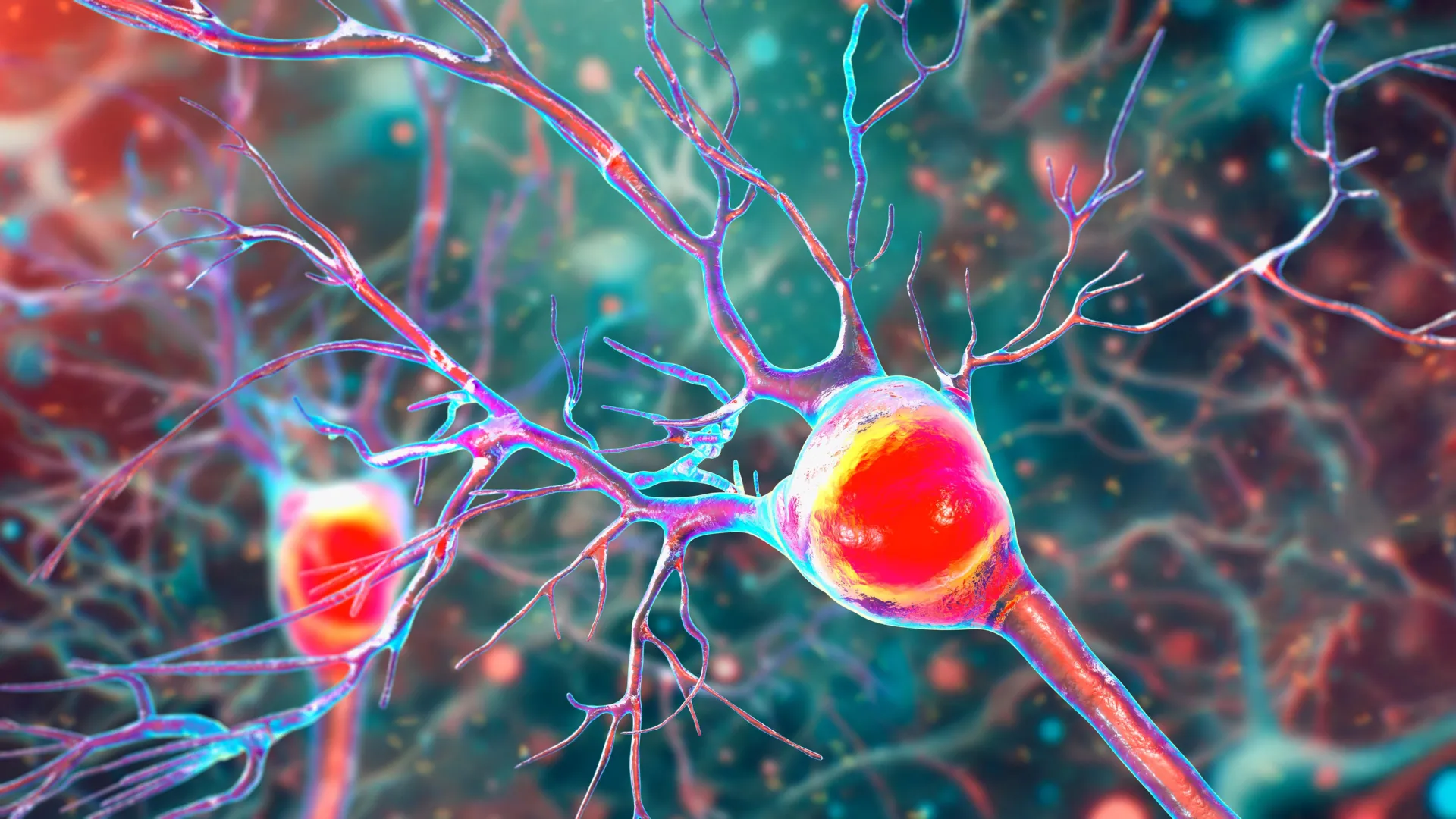
The cerebellum’s ability to manage complex tasks—such as dancing, walking in a straight line, or throwing a ball—depends entirely on the health of Purkinje neurons. These are among the largest and most metabolically active cells in the human brain, characterized by a vast, tree-like structure of dendrites that receive signals from thousands of other neurons.
"Inside the cerebellum are special cells called Purkinje neurons," Tiwari-Woodruff said. "These large, highly active cells help coordinate smooth, precise movements. They are essential for balance and fine motor skills." Because these cells have such high energy demands, they are uniquely vulnerable to mitochondrial impairment. In MS and related neurodegenerative disorders, the gradual death of these cells leads to ataxia—a condition characterized by a lack of voluntary coordination of muscle movements.
The UCR team analyzed postmortem brain tissue from MS patients, comparing it to healthy donor tissue. The findings were stark. The Purkinje cells in MS patients exhibited significantly fewer branches, extensive myelin loss, and clear signs of mitochondrial failure. This lack of energy supply precedes the actual death of the cell, suggesting a window of time where medical intervention might still be possible if the energy supply can be restored.
Insights from the EAE Mouse Model
Because human postmortem tissue only provides a snapshot of the end stages of the disease, the researchers utilized a mouse model known as experimental autoimmune encephalomyelitis (EAE). This model replicates many of the clinical and pathological features of MS, allowing scientists to track the progression of the disease in real-time.
By monitoring the mice over several weeks, the researchers were able to establish a clear chronology of cellular decline. They found that myelin breakdown occurs early in the disease process, followed shortly by the onset of mitochondrial dysfunction. Interestingly, while the "power failure" in the cells began early, the actual death of the Purkinje neurons happened much later, as the disease transitioned into a more severe, chronic phase.
"The remaining neurons don’t work as well because their mitochondria start to fail," Tiwari-Woodruff noted. "The loss of energy in brain cells seems to be a key part of what causes damage in MS." This finding is pivotal because it suggests that the neurological symptoms of MS are not just the result of "broken wires" (demyelination), but of "failing batteries" (mitochondria).
A New Frontier: Targeting Mitochondria as a Therapeutic Strategy
The implications of this research for the pharmaceutical industry and clinical practice are profound. Currently, most FDA-approved treatments for MS are immunomodulatory, meaning they work by suppressing the immune system to prevent new inflammatory attacks. While these "disease-modifying therapies" (DMTs) are effective at reducing the frequency of relapses in the early stages of the disease, they have historically struggled to slow down the long-term progression of disability in secondary progressive MS (SPMS).
The UCR study suggests that to truly halt the progression of MS, researchers must look beyond the immune system and focus on neuroprotection and metabolic support. "Targeting mitochondrial health may represent a promising strategy to slow or prevent neurological decline and improve quality of life for people living with MS," Tiwari-Woodruff stated.
Potential future treatments could involve:
- Bioenergetic Support: Developing drugs that boost COXIV levels or enhance the efficiency of the electron transport chain in neurons.
- Early Intervention: Identifying biomarkers of mitochondrial stress before Purkinje cells are lost, allowing for treatment during the "window of opportunity."
- Remyelination Therapies: Combining mitochondrial support with treatments that encourage the body to repair the myelin sheath, thereby reducing the metabolic strain on the underlying axons.
Future Research and the Search for Cellular Synergy
The UCR research team is not stopping at Purkinje cells. They are currently expanding their investigation to determine if mitochondrial failure is a localized phenomenon or if it extends to other cell types within the cerebellum. This includes oligodendrocytes, the cells responsible for creating myelin, and astrocytes, which provide structural and nutritional support to the entire brain.
"To answer this, one of our ongoing research projects is focused on studying mitochondria in specific types of brain cells in the cerebellum," Tiwari-Woodruff said. Understanding how these different cells interact during the energy crisis of MS could lead to a "cocktail" approach to treatment—one that calms the immune system, repairs myelin, and boosts cellular energy simultaneously.
This holistic approach is particularly vital for addressing the symptoms of balance and coordination loss, which are often the most life-altering for patients. While a patient may be able to manage sensory numbness or occasional fatigue, the loss of independent mobility due to cerebellar ataxia often marks a transition toward a significantly lower quality of life.
The Global Burden and the Case for Scientific Funding
Multiple sclerosis is not only a medical challenge but an economic one. In the United States alone, the total economic burden of MS—including healthcare costs and lost productivity—is estimated to exceed $85 billion annually. As the population ages and the prevalence of progressive forms of MS increases, the need for breakthroughs that preserve function is more urgent than ever.
Professor Tiwari-Woodruff emphasized that continued progress depends heavily on sustained public and private investment in medical research. "Cutting funding to science only slows progress when we need it most," she said, highlighting the role of the National Multiple Sclerosis Society in supporting this specific study.
The research was a collaborative effort, involving a diverse team of scientists including Shane Desfor, Micah Feria, Maria T. Sekyia, Marvellous Osunde, Sandhya Sriram, Saima Noori, Wendy Rincón, and Britany Bello. The use of tissue samples from the National Institutes of Health’s NeuroBioBank and the Cleveland Clinic underscores the importance of large-scale infrastructure in modern neuroscience.
As the scientific community digests the findings of the paper "Decreased mitochondrial activity in the demyelinating cerebellum of progressive multiple sclerosis and chronic EAE contributes to Purkinje cell loss," the focus shifts to the next phase of drug development. By identifying the specific metabolic vulnerabilities of the cerebellum, the UCR team has provided a new map for navigating the complexities of MS, offering hope that the "powerhouses" of the brain can be saved before the lights go out.