
The research is a cornerstone of the Health and Aging Brain Study-Health Disparities (HABS-HD), a comprehensive, multi-university collaboration led by the University of North Texas Health Science Center and supported by the National Institute on Aging (NIA). As Alzheimer’s disease remains the most common cause of dementia worldwide, affecting millions of families and placing an immense burden on healthcare systems, the drive for precise, early-stage biomarkers has never been more urgent.
The Science of Tau and the Search for Clinical Thresholds
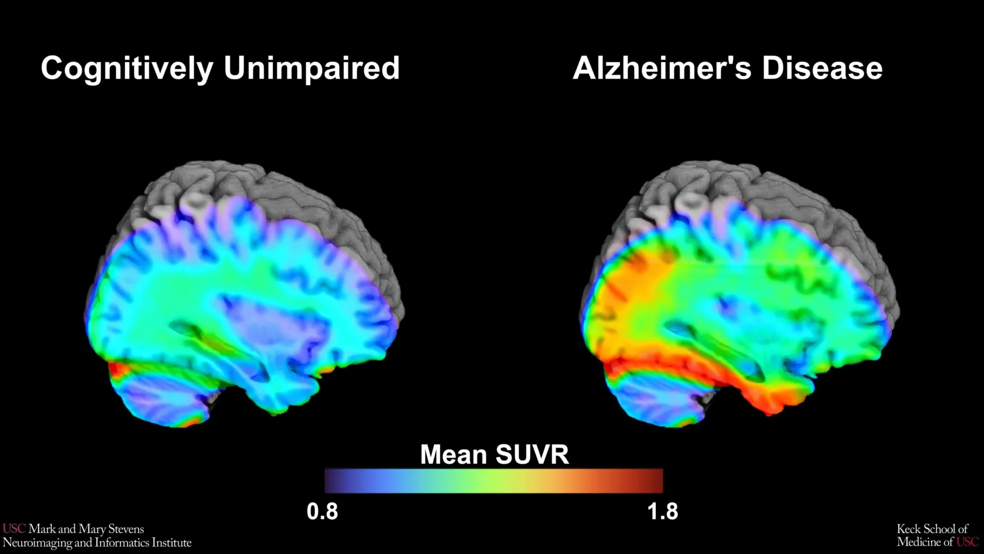
Alzheimer’s disease is characterized by the accumulation of two primary abnormal proteins in the brain: amyloid-beta, which forms plaques between neurons, and tau, which forms tangles inside the neurons. While amyloid plaques often appear years before clinical symptoms manifest, the spread of tau tangles is more closely correlated with the actual onset of memory loss and cognitive impairment.
To visualize these proteins in living patients, researchers utilize Positron Emission Tomography (PET) scans. The USC-led study employed an advanced radioactive tracer known as 18F-PI-2620. This tracer is designed to bind specifically to tau protein deposits, allowing the PET scan to highlight areas of accumulation. The central challenge for the medical community has been determining exactly how much tau constitutes a "positive" diagnosis for Alzheimer’s—a threshold known as a "cut-point."
By studying over 675 older adults from the HABS-HD cohort, the research team sought to identify the optimal brain signal that distinguishes individuals with clinically relevant biological markers of AD from those experiencing normal, age-related changes. The establishment of these cut-points provides a binary metric—positive or negative—that can help clinicians decide whether a patient’s tau levels are high enough to suggest early-stage Alzheimer’s or related neurodegenerative conditions.
Methodology and the Role of the Medial Temporal Lobe
The study’s methodology involved a rigorous comparison of tau PET scans between participants who were classified as cognitively impaired and those who were not, based on a battery of standardized cognitive tests. The researchers focused their analysis on the medial temporal lobe (MTL), a region located deep within the brain that is critical for memory formation and is typically the first area affected by tau pathology in Alzheimer’s disease.
The team discovered that when tau levels in the MTL exceeded a specific threshold, it served as a powerful indicator of cognitive impairment. However, this finding came with a critical caveat: the tau cut-point was most effective only when another abnormal protein, amyloid, was also present. This "dual-protein" requirement underscores the complex interplay between amyloid and tau in the progression of Alzheimer’s.
"Our tau cut-point was able to distinguish whether study participants had cognitive impairment—but only when another abnormal protein, amyloid, was also present in those with cognitive impairment, and only in Hispanic and non-Hispanic White participants," noted senior author Meredith N. Braskie, PhD, assistant professor of neurology at the Keck School of Medicine.
Addressing Health Disparities in Neuroimaging
One of the most striking aspects of the study is its focus on diversity. Historically, the vast majority of Alzheimer’s research has been conducted on non-Hispanic White populations, leading to diagnostic tools and treatment protocols that may not be universally applicable. The HABS-HD initiative was specifically designed to correct this imbalance by recruiting a diverse pool of participants.
The study found that while the tau cut-points were highly effective for Hispanic and non-Hispanic White participants, they did not perform as expected for non-Hispanic Black participants. This discrepancy suggests that cognitive decline in Black populations may be driven by different or additional factors, such as vascular issues, co-pathologies, or social determinants of health that are not captured by current tau imaging benchmarks.
Lead author Victoria R. Tennant, a PhD candidate in USC’s Neuroscience Graduate Program, emphasized the importance of these findings. "The limited reliability of tau as an indicator of cognitive impairment in non-Hispanic Black participants highlights the need for more diverse populations in research and for future studies to examine both biological and social determinants of Alzheimer’s disease," Tennant stated.
Chronology of Alzheimer’s Diagnostic Evolution
The development of the tau cut-point is the latest milestone in a decades-long effort to move Alzheimer’s diagnosis from post-mortem examination to "in-vivo" (in the living) detection.
- 1906: Dr. Alois Alzheimer first describes the plaques and tangles in the brain of a deceased patient.
- 1980s-1990s: Diagnosis relies almost entirely on clinical observation and the exclusion of other dementias.
- 2004: The first amyloid PET tracer (Pittsburgh Compound B) is developed, allowing researchers to see plaques in living brains for the first time.
- 2013: The FDA approves the first tau PET tracer, opening the door for researchers to track the progression of tangles.
- 2020-Present: Large-scale studies like HABS-HD begin focusing on "biomarker-based" diagnosis, where Alzheimer’s is defined by the presence of proteins rather than just symptoms.
- 2024: The USC study establishes the 18F-PI-2620 tau cut-point, providing a specific metric for clinical and research applications in diverse groups.
Supporting Data and Statistical Significance
The study utilized a sophisticated statistical approach to ensure the reliability of the cut-points. By using Receiver Operating Characteristic (ROC) curves, the researchers were able to determine the "Area Under the Curve" (AUC), a measure of how well the tau levels could predict cognitive status.
In Hispanic and non-Hispanic White participants, the presence of elevated tau in the medial temporal lobe, combined with amyloid positivity, showed a high sensitivity and specificity for identifying cognitive impairment. Conversely, in the non-Hispanic Black cohort, the correlation was significantly weaker, suggesting that the "amyloid-tau" model of Alzheimer’s may not fully explain the clinical presentation of the disease in all ethnic and racial groups.
This data is crucial for the design of future clinical trials. If a drug is designed to clear tau from the brain, researchers need a reliable way to identify which participants actually have high tau levels to begin with. Without accurate cut-points tailored to diverse populations, clinical trials run the risk of excluding those who might benefit most or including those whose cognitive decline is not primarily driven by tau.
Institutional and Collaborative Responses
The findings have been met with enthusiasm and a call for further investigation from the broader scientific community. Arthur W. Toga, PhD, director of the Stevens INI, highlighted the broader implications of the HABS-HD project.
"This type of imaging is critical for understanding who is at risk and how the disease develops," Toga said. "These findings are just the latest to come from HABS-HD, which is the most comprehensive study of Alzheimer’s disease and related dementias in diverse communities. HABS-HD has already produced key findings related to ethnic variations in AD biomarkers, the influences of social determinants on cognitive health, and vascular contributions to dementia."
The collaboration involved experts from across the United States, including the Alzheimer’s Therapeutic Research Institute at USC, the Waisman Center at the University of Wisconsin-Madison, Washington University School of Medicine in St. Louis, and the University of California, San Francisco. This multi-institutional support underscores the consensus that Alzheimer’s research must move toward a more inclusive and personalized model.
Broader Impact and Future Clinical Implications
The identification of these new benchmarks has immediate implications for both clinical practice and drug development. As the medical field enters the era of disease-modifying therapies—such as recently approved monoclonal antibodies that target amyloid—the ability to accurately stage a patient’s disease through tau imaging becomes paramount.
- Personalized Medicine: Clinicians may eventually use these cut-points to tailor treatments. For instance, a patient who is "tau-positive" according to the new benchmark might be a candidate for different therapies than one who is "amyloid-positive" but "tau-negative."
- Trial Recruitment: By providing a clear threshold for "tau positivity," the study allows for more precise recruitment in clinical trials, ensuring that the right patients are receiving the right experimental treatments.
- Public Health Policy: The discovery that current biomarkers may not be as effective for Black populations will likely prompt a shift in how public health resources are allocated for dementia screening and research in underserved communities.
- Social Determinants of Health: The study reinforces the theory that biological markers are only one piece of the puzzle. Researchers are now looking more closely at how factors like chronic stress, diet, education, and cardiovascular health contribute to brain aging in different communities.
Conclusion: A New Frontier in Neuroimaging
The work conducted by the Stevens INI and the HABS-HD collaborators marks a pivotal step toward a more equitable and accurate understanding of Alzheimer’s disease. By defining a tau cut-point using the 18F-PI-2620 tracer, the team has provided a new tool for the global effort to diagnose and treat dementia.
However, the study also serves as a sobering reminder of the work that remains. The divergence in results for non-Hispanic Black participants highlights a "missing link" in current neurobiological models. As the scientific community continues to unravel the mysteries of the human brain, the focus must remain on ensuring that every breakthrough, benchmark, and biomarker is applicable to all, regardless of ethnicity or background. The goal is not just a better understanding of Alzheimer’s, but better outcomes and more personalized care for every community affected by this devastating disease.