The findings arrive at a critical juncture in Alzheimer’s research, as the medical community shifts its focus toward precision medicine and the inclusion of diverse demographic groups in clinical trials. By utilizing a sophisticated imaging tracer and analyzing a large, diverse cohort of over 675 older adults, the USC-led team has provided a new lens through which researchers can view the progression of the disease. However, the study also highlights a persistent challenge in neuroimaging: the established benchmarks did not translate effectively to non-Hispanic Black participants, suggesting that the biological or environmental drivers of cognitive decline may differ significantly across racial and ethnic lines.
The Science of Tau and the Mechanics of the New Benchmark
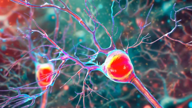
To understand the significance of this study, it is necessary to examine the dual-pathology nature of Alzheimer’s disease. For decades, the "amyloid cascade hypothesis" dominated the field, focusing on the buildup of amyloid-beta plaques between neurons. However, while amyloid appears early in the disease progression, it is the accumulation of tau protein—which forms "tangles" inside neurons—that is more closely correlated with the actual onset of memory loss and cognitive decline.
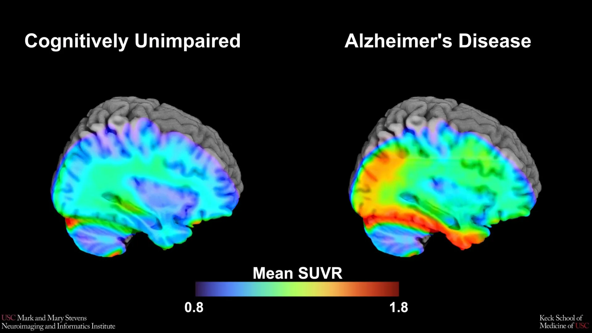
The research team employed an advanced imaging technique known as tau Positron Emission Tomography (PET). This process involves injecting a small amount of a radioactive tracer into the bloodstream. The tracer travels to the brain and binds specifically to tau protein deposits. For this study, the team used a next-generation tracer called 18F-PI-2620. This specific tracer is known for its high affinity and selectivity, allowing researchers to visualize tau accumulation with unprecedented clarity.
By analyzing the scans of 675 participants from the HABS-HD study, the researchers sought to establish "tau cut-points." In medical terms, a cut-point is a specific threshold or numerical value that separates one group from another—in this case, distinguishing individuals with clinically relevant biological markers of Alzheimer’s from those who are experiencing normal, age-related brain changes.
Identifying the Medial Temporal Lobe as a Key Indicator
The study’s findings pinpointed the medial temporal lobe as the primary region for establishing these benchmarks. Located deep within the brain, the medial temporal lobe includes the hippocampus and is essential for the formation of new memories. It is also one of the first areas to be affected by tau pathology in the early stages of Alzheimer’s.
The researchers discovered that when tau levels in the medial temporal lobe exceeded a specific threshold, it served as a robust indicator of cognitive impairment. This finding was particularly strong when paired with the presence of amyloid plaques. This "dual-positivity"—having both high amyloid and high tau—is increasingly seen as the definitive biological signature of Alzheimer’s disease.
"Our tau cut-point was able to distinguish whether study participants had cognitive impairment—but only when another abnormal protein, amyloid, was also present in those with cognitive impairment, and only in Hispanic and non-Hispanic White participants," explained senior author Meredith N. Braskie, PhD, an assistant professor of neurology at the Keck School of Medicine.
A Crucial Discovery in Health Disparities
While the benchmark proved effective for Hispanic and non-Hispanic White participants, the results for non-Hispanic Black participants revealed a significant gap in current neuroimaging models. In this group, the tau cut-point did not accurately predict cognitive impairment. This discrepancy is one of the study’s most vital contributions to the field, as it underscores the limitations of "one-size-fits-all" diagnostic tools.
Lead author Victoria R. Tennant, a PhD candidate in USC’s Neuroscience Graduate Program, noted that the limited reliability of tau as a universal indicator for all races highlights the complexity of the disease. "The limited reliability of tau as an indicator of cognitive impairment in non-Hispanic Black participants highlights the need for more diverse populations in research and for future studies to examine both biological and social determinants of Alzheimer’s disease," Tennant stated.
Experts suggest that in non-Hispanic Black populations, other factors may play a more prominent role in cognitive decline. These could include vascular contributions to dementia—such as high blood pressure or diabetes—as well as social determinants of health, including chronic stress, environmental factors, and historical disparities in healthcare access. The USC study suggests that for these populations, a different set of biomarkers or a different combination of imaging metrics may be required to achieve the same diagnostic accuracy.
Chronology and Context: The Evolution of Alzheimer’s Diagnostics
The development of this new benchmark is the latest step in a decades-long effort to move Alzheimer’s diagnosis from the autopsy table to the living clinic.
- The 1980s-1990s: Alzheimer’s could only be definitively diagnosed post-mortem by examining brain tissue for plaques and tangles.
- Early 2000s: The advent of Amyloid PET imaging allowed doctors to see plaques in living patients, but many people with plaques never developed dementia.
- 2010s: Tau PET imaging was developed, providing a closer link to actual cognitive symptoms.
- 2020-Present: Researchers focus on "cut-points" and "thresholds" to determine exactly when a protein buildup becomes a clinical problem.
The HABS-HD project, which provided the data for this study, was specifically designed to address the historical lack of diversity in these research milestones. Traditionally, over 90% of Alzheimer’s research participants have been of European descent. HABS-HD is the first major study to recruit large, representative cohorts of Black and Hispanic Americans, who are statistically at higher risk for Alzheimer’s but have been chronically underrepresented in clinical trials.
Supporting Data and Statistical Significance
The study’s reliance on 675 participants provides a high level of statistical power. In the field of neuroimaging, where individual scans are expensive and time-consuming, a cohort of this size is substantial. By comparing those with cognitive impairment (based on standardized neuropsychological testing) against cognitively healthy controls, the researchers were able to calculate the sensitivity and specificity of the 18F-PI-2620 tracer.
Data from the Alzheimer’s Association indicates that Black Americans are approximately twice as likely to have Alzheimer’s or other dementias as White Americans, while Hispanics are about 1.5 times as likely. Despite this, the biological pathways are not always identical. The USC study’s data suggests that while tau is a major player in the disease for all groups, its role as a primary diagnostic marker may be more variable than previously thought.
Institutional Collaboration and Official Responses
The study was a massive undertaking involving nearly 30 researchers from several top-tier institutions, including the Alzheimer’s Therapeutic Research Institute (ATRI), the University of Wisconsin-Madison, Washington University in St. Louis, and UC San Francisco.
Arthur W. Toga, PhD, director of the Stevens INI, emphasized the importance of the institute’s role in this global effort. "This type of imaging is critical for understanding who is at risk and how the disease develops," Toga said. "These findings are just the latest to come from HABS-HD, which is the most comprehensive study of Alzheimer’s disease and related dementias in diverse communities. We hope this work will lead to more personalized care and better outcomes for all communities."
The research was supported by extensive funding from the National Institutes of Health (NIH), reflecting the federal government’s commitment to solving the Alzheimer’s crisis. Grants from the National Institute on Aging and the Office of the Director provided the necessary infrastructure for the high-cost PET imaging and the recruitment of diverse study participants.
Broader Impact and Future Implications for Clinical Trials
The establishment of a tau cut-point has immediate implications for the pharmaceutical industry. Currently, several new Alzheimer’s drugs, such as Leqembi (lecanemab) and Kisunla (donanemab), target the removal of amyloid from the brain. However, clinical trials for these drugs often use tau levels to screen participants, as patients with too much or too little tau may not respond to the treatment in the same way.
By refining the tau benchmark, the USC study provides drug developers with a more accurate tool for selecting trial participants. If researchers can accurately identify "tau-positive" individuals in Hispanic and White populations using the 18F-PI-2620 tracer, they can better predict who will benefit from emerging therapies.
Furthermore, the study serves as a "call to action" for the scientific community to investigate why these markers differ in Black populations. This could lead to the discovery of new therapeutic targets that are more effective for Black patients, such as treatments focusing on neuroinflammation or vascular health rather than just protein accumulation.
Conclusion: Toward a Personalized Approach to Brain Health
The research conducted by the Stevens INI and the HABS-HD collaboration represents a pivotal shift in the fight against Alzheimer’s. By moving away from a monolithic view of the disease and toward a nuanced understanding of how it affects different populations, the team at USC is paving the way for a future where Alzheimer’s is not just diagnosed, but predicted and prevented.
As the global population ages, the burden of Alzheimer’s is expected to grow. Benchmarks like the tau cut-point identified in this study are essential tools that will eventually allow clinicians to interpret PET scans with the same precision used in reading cholesterol levels or blood pressure. While more work remains to be done to ensure these tools work for every individual regardless of their background, this study marks a definitive step toward a more equitable and effective era of neurological care.