The identification of this benchmark is particularly timely as the field of neurology moves toward more personalized and inclusive diagnostic frameworks. For decades, Alzheimer’s research has been criticized for its lack of diversity, often relying on data from predominantly White, high-income populations. The new USC study addresses this gap by utilizing a diverse cohort to determine how biological markers of the disease manifest across different ethnic and racial groups, providing a more granular understanding of the disease’s progression.
The Evolution of Tau PET Imaging and Diagnostic Biomarkers
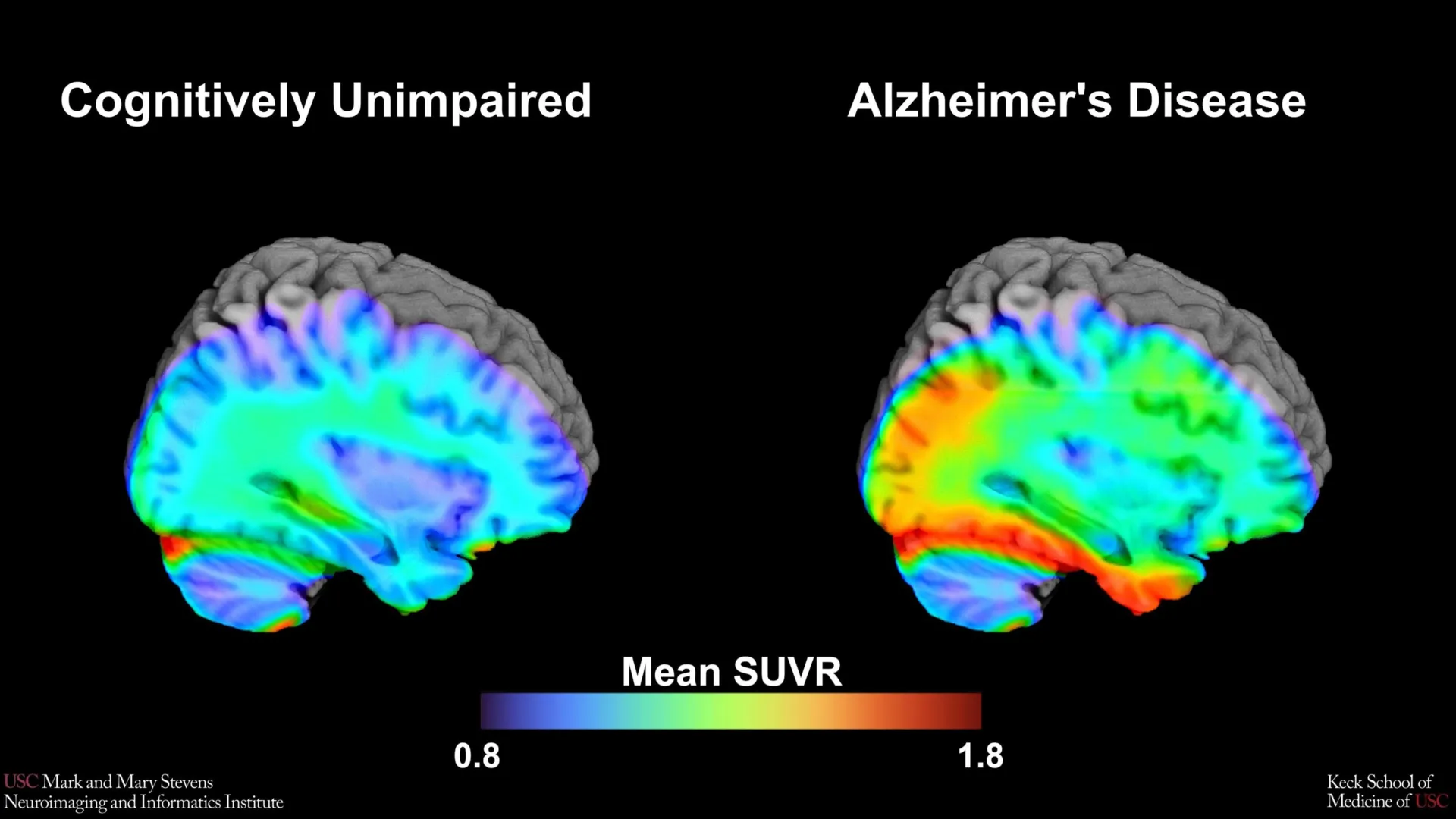
At the heart of this study is an advanced neuroimaging technique known as tau Positron Emission Tomography (PET). Unlike traditional MRI scans that show the structure of the brain, PET scans allow researchers to visualize metabolic processes and the accumulation of specific proteins. In the context of Alzheimer’s disease, the focus is often on two primary "villains": amyloid-beta plaques and tau tangles. While amyloid plaques are often the first sign of the disease, appearing years before symptoms manifest, tau tangles are more closely correlated with actual cognitive decline and the death of neurons.
To visualize these tau tangles, researchers use a radioactive tracer—in this specific study, a newer agent called 18F-PI-2620. This tracer is injected into the bloodstream and binds to tau proteins in the brain, "lighting up" on the PET scan to show exactly where the protein has accumulated. By analyzing these scans from over 675 older adults participating in the HABS-HD study, the USC team sought to establish "tau cut-points." These cut-points act as a diagnostic threshold; if the amount of tau protein in a specific region of the brain exceeds this numerical value, it serves as a biological signal that the individual is likely experiencing early-stage Alzheimer’s or is at high risk for imminent cognitive impairment.
Methodology: Establishing the Medial Temporal Lobe Benchmark
The research team focused their analysis on the medial temporal lobe, a region deep within the brain that is essential for memory formation and is typically one of the first areas affected by tau pathology. By comparing the tau PET scans of study participants who were already experiencing cognitive impairment with those who were aging normally (based on a battery of standardized cognitive tests), the researchers were able to pinpoint a specific level of tau accumulation that distinguished the two groups.
The study found that when tau levels in the medial temporal lobe exceeded the established cut-point, it served as a robust indicator of AD-related cognitive impairment. However, the data revealed a critical caveat: this benchmark was most effective when another abnormal protein, amyloid-beta, was also present. This "amyloid-dependent" tau accumulation reinforces the prevailing scientific theory that amyloid acts as a catalyst, potentially "triggering" the spread of tau throughout the brain, which then leads to the destruction of cognitive function.
"Establishing a cut-point in this region using 18F-PI-2620 marks an important step toward defining tau positivity for both research and clinical applications," stated lead author Victoria R. Tennant, a PhD candidate in USC’s Neuroscience Graduate Program. Tennant noted that while the medial temporal lobe has long been a region of interest, having a standardized numerical threshold allows for more objective comparisons across different clinical trials and patient populations.
Addressing the Disparity in Diagnostic Efficacy
One of the most significant and sobering findings of the study was the variation in how well the tau benchmark performed across different demographic groups. While the tau cut-point was highly effective in identifying cognitive impairment in Hispanic and non-Hispanic White participants, it did not perform as expected in non-Hispanic Black participants.
In the Black participant group, the correlation between this specific tau threshold and cognitive impairment was significantly weaker. This suggests that the biological drivers of cognitive decline may differ across racial and ethnic lines. Senior author Meredith N. Braskie, PhD, an assistant professor of neurology at the Keck School of Medicine, emphasized that this finding does not mean Black individuals are not affected by Alzheimer’s, but rather that other underlying factors—such as vascular health issues, different protein expressions, or social determinants of health—might be playing a more dominant role in their cognitive decline.
"In non-Hispanic Black participants, the tau cut-point did not perform as expected," Braskie explained. "This suggests that other pathologies or conditions may be driving cognitive decline in this group. Our study is an important step toward better understanding how tau relates to cognition in diverse populations and has important implications for future clinical trials that aim to target tau."
The Role of HABS-HD in Modern Neurology
The Health and Aging Brain Study-Health Disparities (HABS-HD) is the most comprehensive study of its kind, specifically designed to investigate why certain populations are disproportionately affected by Alzheimer’s and related dementias. Statistics from the Alzheimer’s Association indicate that Black Americans are about twice as likely, and Hispanics about 1.5 times as likely, to have Alzheimer’s or other dementias compared to older White Americans. Despite these statistics, these groups have historically been underrepresented in the clinical trials that determine how the disease is diagnosed and treated.
Arthur W. Toga, PhD, director of the Stevens INI, highlighted the broader mission of the institute and the HABS-HD collaboration. "This type of imaging is critical for understanding who is at risk and how the disease develops," Toga said. "HABS-HD has already produced key findings related to ethnic variations in AD biomarkers, the influences of social determinants on cognitive health, and vascular contributions to dementia. We hope this work will lead to more personalized care and better outcomes for all communities."
The data produced by HABS-HD allows researchers to look beyond the "one-size-fits-all" model of medicine. By identifying that tau PET scans may need to be interpreted differently depending on a patient’s background, the USC team is paving the way for "precision medicine" in neurology.
Historical Context and the Changing Landscape of Alzheimer’s Research
To understand the weight of these findings, one must look at the history of Alzheimer’s diagnostics. For most of the 20th century, a definitive diagnosis of Alzheimer’s could only be made post-mortem through an autopsy. It wasn’t until the early 2000s that the first amyloid PET tracers were approved, allowing doctors to see the disease in living patients. Tau PET imaging is an even more recent development, with the first tracers receiving FDA approval only within the last several years.
The shift from observing symptoms (like memory loss) to identifying biological markers (like tau levels) represents a paradigm shift. However, as the USC study suggests, the "biological markers" themselves are influenced by a complex interplay of genetics and environment. The fact that the tau benchmark failed to accurately predict impairment in Black participants highlights the "missing data" in the history of neurology. It suggests that current diagnostic tools, while technologically advanced, may still be calibrated to a narrow demographic, potentially leading to misdiagnosis or delayed treatment for minority patients.
Future Implications for Clinical Trials and Treatment
The implications of this study extend directly into the development of new drugs. Recent FDA-approved treatments for Alzheimer’s, such as lecanemab, focus on clearing amyloid from the brain. However, many researchers believe that the next generation of "blockbuster" drugs will need to target tau to truly stop cognitive decline.
If clinical trials for tau-targeting drugs use a universal cut-point to enroll participants, they may inadvertently exclude Black patients who are suffering from cognitive decline but do not meet the "White-calibrated" tau threshold. Conversely, they might enroll patients for whom tau is not the primary driver of their symptoms, leading to failed trials and skewed data. By refining these benchmarks and acknowledging their limitations, the USC study provides a roadmap for more inclusive and effective clinical trial design.
Collaborative Efforts and Funding
The success of this study was made possible through a massive collaborative effort involving experts from across the United States. In addition to the lead researchers at USC, the study included contributors from the Alzheimer’s Therapeutic Research Institute (ATRI), the University of Wisconsin-Madison, the University of North Texas Health Science Center, Washington University School of Medicine in St. Louis, and the University of California, San Francisco.
The research was heavily supported by the National Institute on Aging (NIA) of the National Institutes of Health (NIH), under several grants including R01AG054073 and U19AG078109, as well as the NIH Office of the Director. This level of federal support underscores the national priority of solving the Alzheimer’s crisis, which currently affects over 6 million Americans—a number projected to nearly double by 2050 without significant medical breakthroughs.
Conclusion: A Step Toward Equitable Care
As the medical community continues to unravel the complexities of the human brain, the work being done at the Stevens INI serves as a reminder that scientific progress is inseparable from social equity. The identification of the medial temporal lobe tau cut-point provides a powerful new tool for clinicians, but the discovery of its demographic limitations is perhaps even more vital. It serves as a call to action for the scientific community to investigate the "other pathologies"—be they vascular, inflammatory, or environmental—that contribute to brain aging in diverse communities.
Ultimately, the goal is to ensure that when a patient walks into a clinic concerned about their memory, the diagnostic tools used to evaluate them are as diverse as the population they serve. Through the ongoing work of the HABS-HD and the Keck School of Medicine, the path toward that goal is becoming clearer, one scan at a time.