New findings from a research team at the University of California, Riverside (UCR) have provided a critical breakthrough in understanding why this cerebellar decline occurs. Published in the prestigious journal Proceedings of the National Academy of Sciences (PNAS), the study identifies the failure of mitochondria—the internal "power plants" of the cell—as a primary driver behind the death of Purkinje cells. These specific neurons are the output engines of the cerebellum, and their loss is directly correlated with the progressive mobility issues that define the lived experience of many MS patients. By pinpointing mitochondrial dysfunction as a central mechanism of neurodegeneration, the research opens a new frontier for therapeutic interventions aimed at preserving energy production within the brain.
The Biological Mechanism of Decline: Inflammation and Demyelination
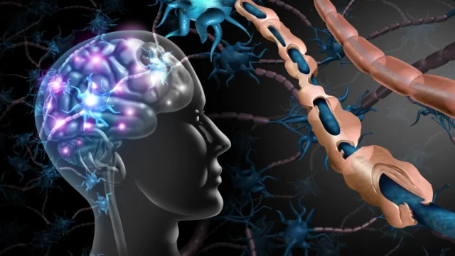
The hallmark of multiple sclerosis is a two-pronged assault on the nervous system: inflammation and demyelination. In a healthy body, nerve fibers are coated in a fatty substance called myelin, which acts as an insulating layer similar to the plastic coating on an electrical wire. This sheath allows electrical impulses to travel rapidly and efficiently between the brain and the rest of the body. In patients with MS, the immune system mistakenly identifies myelin as a foreign threat, leading to its destruction.
When demyelination occurs in the cerebellum, the communication lines responsible for motor coordination begin to fray. However, the UCR study suggests that the damage goes deeper than just the loss of insulation. The research team, led by Seema Tiwari-Woodruff, a professor of biomedical sciences at the UC Riverside School of Medicine, discovered that the inflammatory environment of MS disrupts the internal machinery of the neurons themselves.
"Our study, conducted by my graduate student Kelley Atkinson, proposes that inflammation and demyelination in the cerebellum disrupt mitochondrial function, contributing to nerve damage and Purkinje cell loss," Tiwari-Woodruff explained. The team specifically identified a significant reduction in a protein known as COXIV (Cytochrome c oxidase subunit 4). This protein is essential for the mitochondrial electron transport chain, the process by which cells convert oxygen and nutrients into adenosine triphosphate (ATP), the universal energy currency of life. Without sufficient COXIV, the Purkinje cells experience a catastrophic energy failure, leading to cellular stress and, eventually, programmed cell death.
The Vital Role of Purkinje Cells in Human Mobility
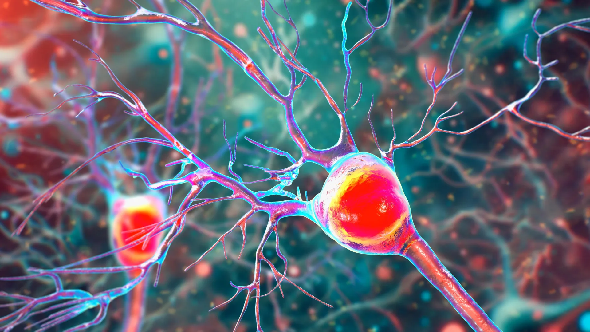
To understand the impact of this research, one must understand the unique role of Purkinje neurons. These are among the largest and most metabolically demanding cells in the human brain. Their intricate, tree-like branching structures—known as dendritic trees—allow them to receive and process a massive amount of sensory information from across the body.
"Inside the cerebellum are special cells called Purkinje neurons," said Tiwari-Woodruff. "These large, highly active cells help coordinate smooth, precise movements—like dancing, throwing a ball, or even just walking. They’re essential for balance and fine motor skills."
In the context of MS, the high energy demands of Purkinje cells make them particularly vulnerable to mitochondrial impairment. When the energy supply fails, these neurons cannot maintain their complex structures. The UCR researchers observed that in the brains of MS patients, the remaining Purkinje cells exhibited fewer branches and significant "blebbing" or thinning of the axons. As these neurons die off, the cerebellum loses its ability to regulate movement, leading to a condition known as ataxia. Ataxia manifests as slurred speech, stumbling, and an inability to perform tasks requiring manual dexterity, such as buttoning a shirt or writing.
Comparative Analysis: Human Tissue and the EAE Mouse Model
The study utilized a comprehensive dual-track methodology to validate its findings. First, the researchers analyzed postmortem cerebellar tissue from individuals who had been diagnosed with secondary progressive MS, a stage of the disease where disability increases steadily. These samples were provided by the National Institutes of Health’s NeuroBioBank and the Cleveland Clinic, allowing the team to observe the end-stage effects of the disease in human subjects.
To observe the progression of the disease in real-time, the team also employed the experimental autoimmune encephalomyelitis (EAE) mouse model. EAE is a widely recognized surrogate for MS in laboratory settings, as it mimics the immune-mediated demyelination and neurodegeneration seen in humans. By tracking the mice from the onset of symptoms through the chronic phase of the disease, the researchers were able to establish a clear timeline of biological failure.
The results from the EAE model mirrored the human data with striking accuracy. The mice showed early signs of myelin breakdown, followed closely by a decline in mitochondrial activity. As the disease transitioned into a chronic state, the loss of Purkinje cells accelerated, coinciding with the onset of severe motor deficits. "The loss of energy in brain cells seems to be a key part of what causes damage in MS," Tiwari-Woodruff noted, emphasizing that while demyelination happens early, the actual death of the neurons is a secondary consequence of the sustained metabolic crisis.
Data and Statistics: The Growing Burden of Multiple Sclerosis
The implications of this research are underscored by the rising prevalence and economic impact of MS. According to the National MS Society, the prevalence of the disease in the United States has nearly doubled in recent decades, with nearly 1 million adults currently living with the diagnosis. Globally, the disease disproportionately affects women, who are diagnosed at a rate three times higher than men.
The economic burden is equally staggering. A 2022 study estimated the total economic cost of MS in the United States at over $85 billion annually, including direct medical costs and indirect costs such as lost productivity and caregiver expenses. Much of this cost is driven by the transition from relapsing-remitting MS to progressive forms of the disease, where mobility loss becomes permanent. Current FDA-approved treatments are largely effective at reducing the frequency of inflammatory "flares," but they have shown limited success in stopping the underlying neurodegeneration that leads to long-term disability. The UCR study provides a potential roadmap for a new class of "neuroprotective" therapies that could fill this gap.
Future Research and Potential Therapeutic Strategies
The identification of mitochondrial failure as a culprit in Purkinje cell death suggests that future treatments could focus on "bioenergetic support." This might involve drugs designed to stabilize the COXIV protein, antioxidants that target the mitochondria to reduce oxidative stress, or metabolic boosters that help neurons maintain ATP production despite the presence of inflammation.
However, the UCR team is not stopping at Purkinje cells. Their ongoing research is investigating whether this "energy crisis" extends to other critical cells in the cerebellum. These include:
- Oligodendrocytes: The cells responsible for creating and repairing the myelin sheath. If their mitochondria fail, the brain loses its ability to remyelinate damaged areas.
- Astrocytes: Star-shaped cells that provide structural support and regulate the chemical environment of the brain. Malfunctioning astrocytes can contribute to a toxic environment that further harms neurons.
"To answer this, one of our ongoing research projects is focused on studying mitochondria in specific types of brain cells in the cerebellum," Tiwari-Woodruff said. "Such research can open the door to finding ways to protect the brain early on—like boosting energy in brain cells, helping them repair their protective myelin coating, or calming the immune system before too much damage is done."
The Critical Role of Scientific Funding
The study was supported by funding from the National Multiple Sclerosis Society, a reminder of the reliance of medical breakthroughs on public and private philanthropy. In the concluding remarks of her report, Professor Tiwari-Woodruff addressed the broader political and social climate surrounding scientific research. She argued that sustained investment is the only way to transform these laboratory findings into bedside treatments.
"Cutting funding to science only slows progress when we need it most," she stated. "Public support for research matters now more than ever."
As the scientific community moves forward, the UCR study stands as a pivotal piece of the puzzle. By shifting the focus from the "insulation" of the nerves to the "engines" that power them, researchers are moving closer to a future where MS is no longer a sentence of progressive physical decline, but a manageable condition where the brain’s vital functions are preserved through targeted, energy-focused medicine. The work of Tiwari-Woodruff, Atkinson, and their colleagues—including Shane Desfor, Micah Feria, and others—represents a significant step toward reclaiming the quality of life for millions of people worldwide.