
Multiple sclerosis (MS) currently affects approximately 2.3 to 2.8 million people globally. While the disease is often associated with lesions in the white matter of the brain and spinal cord, approximately 80% of clinical cases involve significant pathology in the cerebellum. Located at the back of the skull, the cerebellum is responsible for the "fine-tuning" of motor activity. When this region is compromised, patients experience ataxia—a condition characterized by a lack of voluntary coordination of muscle movements that can include gait abnormality, speech changes, and abnormalities in eye movements. The UCR study provides a granular look at why the cerebellum is so vulnerable and how the failure of the "powerhouses of the cell" leads to permanent neurological disability.
The Role of the Cerebellum and Purkinje Neurons
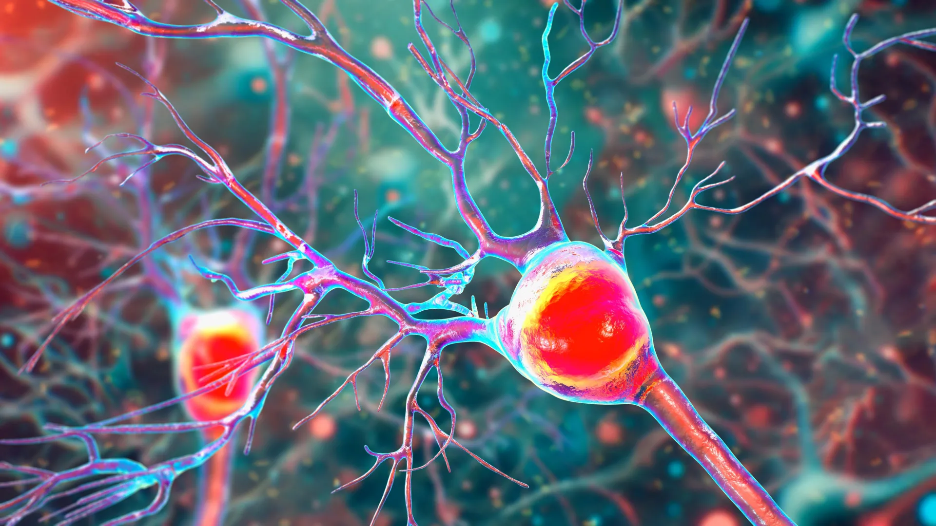
To understand the impact of the UCR findings, it is necessary to consider the unique architecture of the cerebellum. Although it accounts for only about 10% of the brain’s total volume, the cerebellum contains more than half of the brain’s total neurons. Among these, the Purkinje cells are the most distinctive. These are some of the largest neurons in the human brain, characterized by an intricately branched "dendritic tree" that allows them to receive and process a massive amount of inhibitory and excitatory information.
Purkinje cells are the sole output for all motor coordination signals from the cerebellar cortex. If these cells are damaged or lost, the brain’s ability to coordinate smooth, precise movements is essentially severed. Professor Seema Tiwari-Woodruff, a lead author of the study and a professor of biomedical sciences at the UC Riverside School of Medicine, noted that these cells are essential for everything from high-level athletic performance to the simple act of walking in a straight line. The research team found that in MS patients, these neurons undergo a process of "arborization failure," where their branches wither before the cell eventually dies.
Investigating Mitochondrial Dysfunction and COXIV Protein Loss
The central discovery of the UCR research involves the role of mitochondria in the survival of Purkinje cells. Mitochondria are organelles responsible for producing adenosine triphosphate (ATP), the chemical energy currency of the cell. In the context of MS, the research team identified a significant reduction in a specific mitochondrial protein known as COXIV (Cytochrome c oxidase subunit 4).
COXIV is a key component of the mitochondrial respiratory chain. Its reduction signifies a breakdown in the cell’s ability to utilize oxygen to produce energy. The study, spearheaded by graduate student Kelley Atkinson, utilized postmortem cerebellar tissue from individuals who had been diagnosed with secondary progressive MS. By comparing this tissue with samples from healthy donors—sourced from the National Institutes of Health’s NeuroBioBank and the Cleveland Clinic—the researchers were able to observe a direct correlation between demyelination and the loss of COXIV.
Demyelination is the hallmark of MS, where the immune system attacks the myelin sheath, the fatty insulation surrounding nerve fibers. Without myelin, nerve impulses travel more slowly or not at all. However, the UCR study suggests a secondary, perhaps more lethal, effect: the loss of myelin disrupts the metabolic support provided to the axon, forcing the mitochondria to work harder under stress. Eventually, the mitochondria fail, leading to a state of "energy failure" that triggers programmed cell death in the Purkinje neurons.
Evidence from the EAE Mouse Model
To validate their findings and observe the progression of the disease in real-time, the researchers employed the experimental autoimmune encephalomyelitis (EAE) mouse model. EAE is the most commonly used animal model for MS, as it mimics the inflammatory and demyelinating aspects of the human condition.
The longitudinal study of these mice allowed the team to establish a timeline of decay. They observed that myelin breakdown occurs early in the disease cycle, followed closely by a decline in mitochondrial efficiency. While the neurons initially survive the loss of their protective coating, the subsequent energy failure proves fatal. The researchers tracked a steady decline in the number of viable Purkinje cells as the mice’s motor symptoms—such as tail paralysis and limb weakness—intensified.
"The remaining neurons don’t work as well because their mitochondria start to fail," Tiwari-Woodruff explained. This finding is significant because it suggests a window of opportunity for intervention. If the "death of the brain cells tends to happen later," as the study indicates, then therapeutic strategies aimed at boosting mitochondrial health early in the disease could potentially save these neurons and preserve motor function.
Shifting the Therapeutic Paradigm: From Inflammation to Neuroprotection
For decades, the primary strategy for treating MS has focused on immunotherapy—suppressing the immune system to prevent it from attacking the myelin sheath. While these "disease-modifying therapies" (DMTs) have been successful in reducing the frequency of relapses in relapsing-remitting MS, they have been less effective in stopping the slow, steady decline seen in progressive forms of the disease.
The UCR study adds to a growing body of evidence suggesting that neuroprotection—specifically targeting the metabolic health of neurons—must be a co-priority. By identifying COXIV loss as a driver of cell death, the research opens the door for "mitochondrial-centric" therapies. This could include the development of drugs that stabilize the mitochondrial membrane, antioxidants targeted specifically at the mitochondria, or metabolic precursors that help maintain ATP production despite the loss of myelin.
The implications for quality of life are profound. For many MS patients, the transition from relapsing-remitting to secondary progressive MS is marked by a loss of independence due to mobility issues. Protecting the Purkinje cells could mean the difference between a patient remaining ambulatory or requiring a wheelchair.
Broader Research and Future Directions
The UCR team is not stopping at Purkinje cells. The research is expanding to look at how mitochondrial failure affects other cell types within the cerebellum. This includes oligodendrocytes, the cells responsible for creating myelin, and astrocytes, which provide structural and nutritional support to the brain.
If mitochondrial dysfunction is a systemic issue across multiple cell types in the MS brain, the treatment strategy may need to be even more holistic. "Such research can open the door to finding ways to protect the brain early on—like boosting energy in brain cells, helping them repair their protective myelin coating, or calming the immune system before too much damage is done," said Tiwari-Woodruff.
The study also underscores the necessity of high-quality tissue banks and sustained research funding. The use of postmortem human tissue was vital in confirming that the patterns seen in the mouse models were actually occurring in human patients. Tiwari-Woodruff emphasized that public support for science is a prerequisite for these types of breakthroughs, noting that any reduction in funding directly translates to a slowdown in the development of life-changing treatments.
Analysis of Clinical and Economic Impacts
The economic burden of MS is substantial, with costs related to healthcare, lost productivity, and informal care estimated at billions of dollars annually in the United States alone. A significant portion of these costs is driven by the progression of disability. By identifying a specific protein (COXIV) and a specific cell type (Purkinje neurons) as the locus of decline, this research provides a roadmap for more efficient drug development.
Furthermore, the study highlights the importance of the cerebellum in the broader map of MS research. Historically, much of MS research focused on the cerebral cortex and spinal cord. The UCR study’s focus on the "hindbrain" corrects a long-standing oversight, acknowledging that balance and coordination are often the symptoms that most severely impact a patient’s daily life.
As the scientific community moves forward, the focus will likely shift toward clinical trials that combine traditional immunotherapies with new neuroprotective agents. The goal is a "dual-action" treatment plan: one that stops the immune system’s fire while simultaneously repairing and refueling the neurons caught in the blaze.
The UCR study, titled "Decreased mitochondrial activity in the demyelinating cerebellum of progressive multiple sclerosis and chronic EAE contributes to Purkinje cell loss," stands as a pivotal contribution to the field. It provides a clear biological explanation for the "energy gap" in MS and offers a tangible target for future medical innovation. For the millions of people living with MS, these findings represent a significant step toward a future where the diagnosis of a progressive disease does not inevitably lead to the loss of movement and independence.