
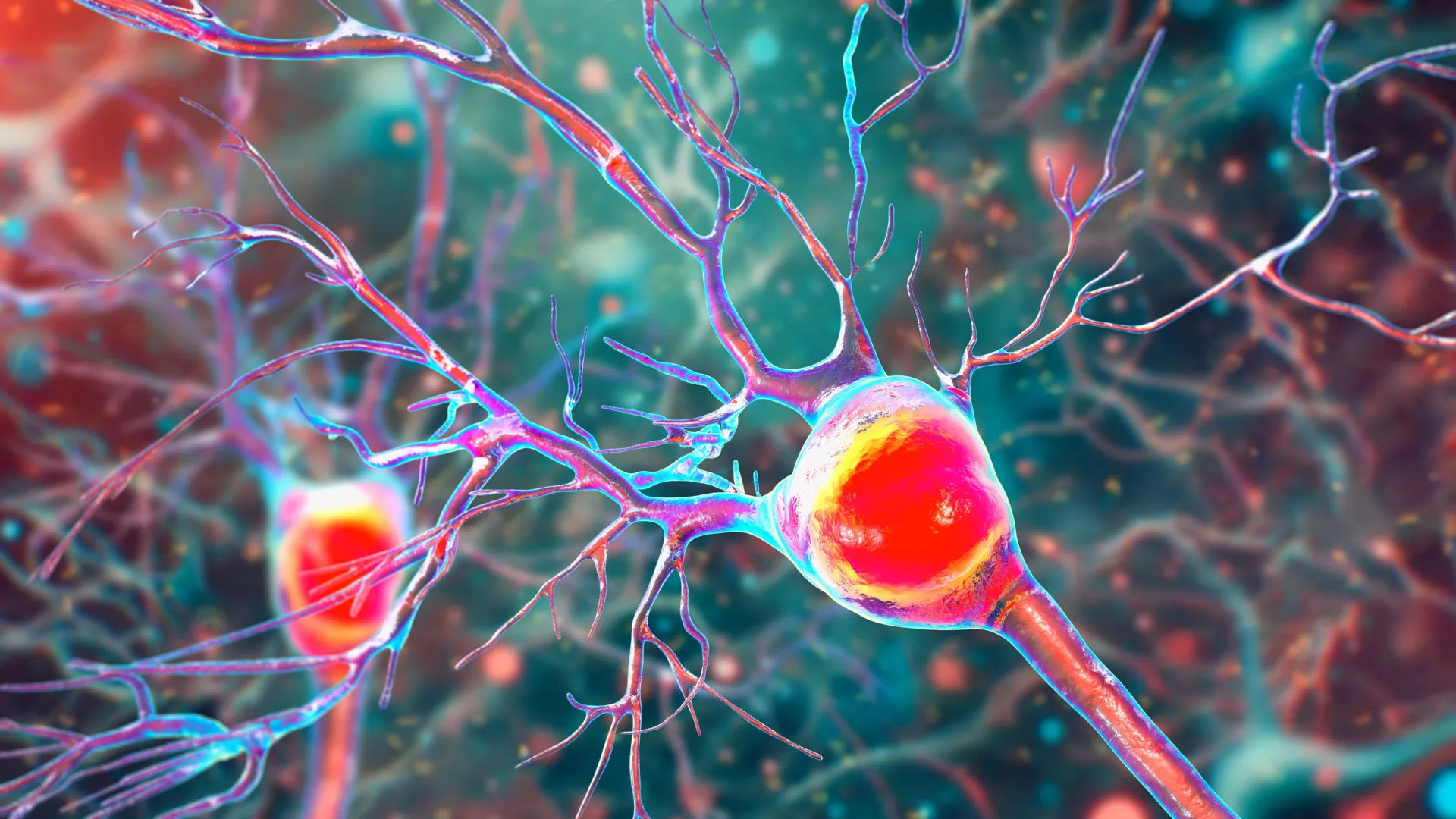
The cerebellum, a region located at the back of the brain, is essential for maintaining balance, posture, and coordinated movement. Clinical data indicates that in roughly 80% of MS cases, this region becomes a focal point for inflammation and tissue damage. When the cerebellum is compromised, patients often experience tremors, an unsteady gait, and a profound loss of fine motor control. The UCR study suggests that the progressive loss of Purkinje cells—large, complex neurons within the cerebellum—is the direct result of an "energy failure" caused by malfunctioning mitochondria.
The Biological Mechanism: When the Powerhouse Fails
Multiple sclerosis is traditionally defined by two hallmark processes: chronic inflammation and demyelination. Demyelination occurs when the immune system mistakenly targets the myelin sheath, the fatty insulation that ensures rapid electrical signaling between neurons. Without this insulation, nerve impulses are slowed or blocked entirely. However, the UCR research team, led by Seema Tiwari-Woodruff, a professor of biomedical sciences, posits that demyelination is only part of a more complex destructive cycle.
The study focused on the role of mitochondria, the organelles responsible for generating adenosine triphosphate (ATP), the chemical energy that powers cellular functions. Through a detailed analysis of both human brain tissue and animal models, the researchers discovered a significant reduction in the mitochondrial protein COXIV. This protein is a critical component of the electron transport chain, which is the final stage of cellular respiration.
"Our study proposes that inflammation and demyelination in the cerebellum disrupt mitochondrial function, contributing to nerve damage and Purkinje cell loss," explained Professor Tiwari-Woodruff. The data suggests that when myelin is lost, the metabolic demands on the underlying axon increase. If the mitochondria are already impaired by inflammatory cytokines, they cannot meet this increased demand, leading to a state of chronic energy deficiency that eventually triggers programmed cell death.
The Critical Role of Purkinje Neurons
To understand the impact of this research, one must understand the unique nature of Purkinje cells. These neurons are among the largest in the human brain and possess an incredibly dense "tree" of branches known as dendrites. They serve as the primary output for the cerebellar cortex, acting as the final checkpoint that coordinates motor signals before they are sent to the rest of the body.
In the healthy brain, Purkinje cells facilitate smooth, precise movements, such as the ability to type on a keyboard, maintain balance while walking on uneven surfaces, or perform the complex motor sequences required for dancing. However, the UCR team found that in MS patients, these cells undergo a visible transformation before they die. The dendritic branches become stunted and sparse, and the cells lose their vital connections to other neurons.
As these neurons disappear, the patient develops ataxia—a neurological sign consisting of a lack of voluntary coordination of muscle movements. The study’s findings indicate that the loss of Purkinje cells is not a random occurrence but is closely tied to the localized failure of mitochondrial energy production in areas where myelin has been stripped away.
Chronology of Neurodegeneration: Tracking Disease Progression
The research team utilized a dual-pronged approach to establish a timeline of how this damage unfolds. First, they analyzed postmortem cerebellar tissue from individuals who had been diagnosed with secondary progressive MS (SPMS), a stage of the disease where disability worsens steadily without distinct periods of relapse and remission. These samples were compared against healthy control tissue provided by the National Institutes of Health’s NeuroBioBank and the Cleveland Clinic.
To observe the disease in real-time, the researchers also employed the experimental autoimmune encephalomyelitis (EAE) mouse model. This model mimics the inflammatory and demyelinating characteristics of human MS, allowing scientists to track cellular changes from the onset of the disease to its chronic stages.
The chronological findings revealed a distinct pattern:
- Early Phase: Inflammation begins in the cerebellum, and the immune system starts attacking the myelin sheath.
- Intermediate Phase: As demyelination progresses, mitochondrial proteins like COXIV begin to decrease. The energy output of the Purkinje cells drops significantly.
- Chronic Phase: The energy-starved Purkinje cells begin to lose their structural integrity. Dendritic branches retreat, and the cells eventually undergo apoptosis (cell death).
- Symptomatic Peak: The physical manifestation of this loss—ataxia and severe mobility issues—becomes permanent as the brain loses its ability to compensate for the missing neurons.
"The remaining neurons don’t work as well because their mitochondria start to fail," Tiwari-Woodruff noted. "These problems start early, but the actual death of the brain cells tends to happen later, as the disease becomes more severe."
Supporting Data and Clinical Implications
The implications of these findings are significant for the future of MS pharmacology. Currently, the majority of FDA-approved disease-modifying therapies (DMTs) focus on the "outside-in" approach—modulating or suppressing the immune system to prevent new inflammatory attacks. While effective at reducing relapses in the early stages of the disease, these treatments have shown limited efficacy in stopping the neurodegeneration that characterizes progressive MS.
The UCR study highlights a need for "inside-out" neuroprotective strategies. By targeting mitochondrial health, researchers hope to provide neurons with the resilience needed to survive inflammatory environments. Potential interventions could include:
- Mitochondrial Boosters: Small molecules designed to enhance the activity of the electron transport chain or stabilize proteins like COXIV.
- Antioxidant Therapy: Reducing the oxidative stress that further damages mitochondria during the inflammatory process.
- Metabolic Support: Providing alternative energy substrates to neurons that are struggling with ATP production.
Data from the study showed that the loss of COXIV was specifically concentrated in demyelinated regions, suggesting that if myelin repair (remyelination) can be paired with mitochondrial support, the survival rate of Purkinje cells could be significantly improved.
Broader Impact on the Scientific Community
The research conducted by Tiwari-Woodruff and her graduate student, Kelley Atkinson, alongside a diverse team of scientists, has been met with interest from the wider neurological community. It reinforces a growing body of evidence that MS is as much a metabolic and degenerative disease as it is an autoimmune one.
The study was supported by funding from the National Multiple Sclerosis Society, an organization that has increasingly prioritized research into the progressive forms of the disease. The findings provide a roadmap for investigating whether similar mitochondrial failures occur in other regions of the brain, such as the motor cortex or the spinal cord, which are also frequently damaged in MS.
Furthermore, the team is now expanding their scope to look at other supporting cells in the brain. They are investigating whether mitochondrial damage extends to oligodendrocytes (the cells that create myelin) and astrocytes (the cells that provide structural and metabolic support to neurons). If these support cells are also suffering from energy failure, it could explain why the brain’s natural repair mechanisms eventually fail in MS patients.
The Necessity of Continued Research Funding
In the wake of these findings, Professor Tiwari-Woodruff emphasized the critical nature of sustained investment in medical science. The complexity of the brain and the sophisticated nature of mitochondrial biology require long-term, high-cost research projects that are often vulnerable to economic shifts.
"Cutting funding to science only slows progress when we need it most," she stated. "Public support for research matters now more than ever."
As the scientific community moves toward a more holistic understanding of multiple sclerosis, the UCR study stands as a pivotal contribution. By identifying the specific "energy crisis" within Purkinje cells, the research offers a new target for drug development, bringing the medical field a step closer to treatments that do not just delay the onset of MS symptoms, but actively protect the brain from the inside out. For the millions of people struggling with the loss of balance and coordination, these insights represent a vital beacon of hope for a future where mobility can be preserved despite a diagnosis of multiple sclerosis.