The research, led by the Lee Kong Chian School of Medicine (LKCMedicine) at NTU, highlights a critical physiological marker that can be identified through routine magnetic resonance imaging (MRI) scans. By recognizing these "clogged drains" before the onset of irreversible neuronal damage, medical professionals may soon have a non-invasive, cost-effective window of opportunity to intervene in the progression of Alzheimer’s disease, which remains the most prevalent cause of dementia worldwide.
The Mechanics of Neurological Waste Removal
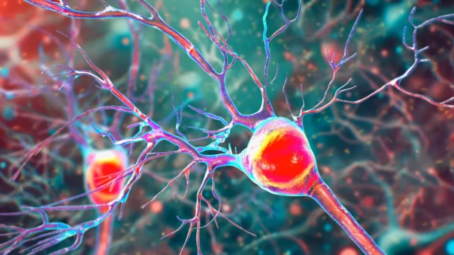
The human brain is an incredibly active organ, consuming a disproportionate amount of the body’s energy and, consequently, producing a significant volume of metabolic waste. To maintain health, the brain utilizes a specialized drainage system. Blood vessels within the brain are enveloped by microscopic channels known as perivascular spaces. These channels act as a plumbing system, facilitating the clearance of interstitial fluid and toxic proteins, most notably beta-amyloid and tau.
In a healthy brain, this drainage system operates efficiently, ensuring that these proteins do not accumulate to toxic levels. However, when the system becomes less effective, these perivascular spaces begin to dilate or enlarge. Under an MRI, these enlarged perivascular spaces (EPVS) become visible as distinct anomalies. The NTU study is among the first to conclusively link the presence and severity of these enlarged spaces to the early biochemical hallmarks of Alzheimer’s disease, particularly within an Asian demographic.
Addressing the Research Gap in Asian Populations
One of the most significant aspects of the NTU Singapore study is its focus on Asian participants. Historically, the vast majority of Alzheimer’s research has been conducted on Caucasian cohorts in North America and Europe. This geographic and ethnic bias in data has long been a concern for global health experts, as the genetic and environmental factors influencing dementia can vary significantly across different populations.
For instance, the apolipoprotein E4 (APOE4) gene is widely recognized as the most significant genetic risk factor for late-onset Alzheimer’s. In Caucasian populations, approximately 50 to 60 percent of Alzheimer’s patients carry this gene. However, Associate Professor Nagaendran Kandiah, the lead investigator of the study and Director of the Dementia Research Centre (Singapore) at LKCMedicine, pointed out that the prevalence of APOE4 among Singaporean dementia patients is less than 20 percent.
This discrepancy underscores the necessity for region-specific research. "Because of these differences, findings in one population may not directly apply to another," Assoc Prof Kandiah explained. The NTU team addressed this gap by examining nearly 1,000 individuals in Singapore, reflecting the nation’s diverse multi-ethnic makeup, including Chinese, Malay, and Indian backgrounds.
Methodology: MRI Analysis and Blood Biomarkers
The study’s rigorous methodology involved a two-pronged approach: advanced neuroimaging and blood-based biochemical analysis. The research team analyzed the MRI scans of nearly 1,000 participants, categorized into those with normal cognitive function and those experiencing mild cognitive impairment (MCI). MCI is often viewed as a transitional stage between the expected cognitive decline of normal aging and the more serious decline of dementia.
To validate the significance of the enlarged perivascular spaces seen on the MRIs, the scientists measured seven specific Alzheimer’s-related biochemicals in the participants’ blood. These markers included various forms of beta-amyloid and tau proteins, which are known to form the plaques and tangles that characterize the Alzheimer’s brain.
The results were striking. The researchers found that participants with MCI were significantly more likely to exhibit enlarged perivascular spaces compared to their cognitively healthy counterparts. Furthermore, the presence of these enlarged spaces was strongly correlated with four of the seven blood biomarkers. This correlation suggests that individuals with "clogged" brain drainage are at a much higher risk of having increased amyloid plaques and tau tangles, even if they have not yet developed full-blown dementia symptoms.
Comparative Sensitivity: EPVS vs. White Matter Damage
Traditionally, clinicians have looked for "white matter hyperintensities"—areas of damage to the brain’s white matter caused by reduced blood flow—as a primary indicator of cognitive decline on MRI scans. While white matter damage is a well-established marker, the NTU study found that enlarged perivascular spaces might be an even more sensitive early warning sign.
Among the participants with mild cognitive impairment, the link between Alzheimer’s-related biochemicals and enlarged perivascular spaces was notably stronger than the link with white matter damage. This suggests that while white matter damage indicates vascular issues, the enlargement of perivascular spaces is a more direct reflection of the brain’s failure to clear the specific proteins that drive Alzheimer’s pathology.
Justin Ong, the study’s first author and a fifth-year student at LKCMedicine, emphasized that this finding is pivotal for early intervention. "Identifying Alzheimer’s sooner gives doctors more time to intervene and potentially slow the progression of symptoms such as memory loss, reduced thinking speed, and mood changes," Ong stated. The research was completed as part of the Scholarly Project module, highlighting the integration of high-level research into the medical curriculum at NTU.
Clinical Reactions and the Synergy of Disease
The medical community has reacted with optimism to the findings. Dr. Rachel Cheong Chin Yee, a Senior Consultant at Khoo Teck Puat Hospital’s Department of Geriatric Medicine, who was not involved in the study, noted the importance of the findings for risk stratification. She suggested that identifying these markers on routine scans could help identify high-risk individuals before symptoms even appear, allowing for lifestyle modifications or early pharmacological treatments.
Furthermore, the study challenges the traditional siloed view of brain diseases. Dr. Chong Yao Feng, a Consultant at the National University Hospital’s Division of Neurology, observed that cerebrovascular disease (issues with blood vessels) and Alzheimer’s disease (a neurodegenerative proteinopathy) have often been treated as separate entities.
"The study’s findings are intriguing as they demonstrate that both diseases do interact in a synergistic manner," Dr. Chong noted. He cautioned that when doctors see markers like enlarged perivascular spaces on an MRI, they should not automatically assume the patient only has a vascular problem. Instead, they must consider the high probability that an underlying Alzheimer’s process is also at play.
Implications for Future Diagnostics and Global Healthcare
The implications of this research extend beyond the laboratory. As the global population ages, particularly in Asia, the socioeconomic burden of Alzheimer’s disease is projected to rise exponentially. Early detection is considered the "holy grail" of dementia care because most current and emerging treatments are most effective in the earliest stages of the disease, before widespread neuronal death occurs.
The fact that these markers can be identified through routine MRI scans is a major advantage. Unlike PET scans or cerebrospinal fluid draws—which can be expensive, invasive, or require specialized facilities—MRIs are already a standard tool in evaluating cognitive decline. By adding the evaluation of perivascular spaces to the standard checklist for radiologists, the medical community could implement a screening process without increasing the financial burden on patients or the healthcare system.
Timeline and Future Directions
The NTU research team has outlined a clear path for the next phase of their investigation. The current study provided a cross-sectional "snapshot" of the link between brain drainage and Alzheimer’s markers. The next step involves a longitudinal study, where the team will track the nearly 1,000 participants over several years.
This follow-up will allow researchers to see exactly how many of the individuals with enlarged perivascular spaces eventually progress to a clinical diagnosis of Alzheimer’s dementia. This data will be crucial for determining the predictive accuracy of EPVS as a diagnostic tool.
If the longitudinal data confirms the current findings, it could lead to a shift in international clinical guidelines for dementia diagnosis. Furthermore, the discovery opens up new avenues for therapeutic research. If "clogged drains" are a primary driver of protein accumulation, then treatments designed to improve the efficiency of the brain’s waste removal system—perhaps through specialized medications or even lifestyle interventions like optimized sleep patterns—could become a new frontier in the fight against Alzheimer’s.
As the study concludes, the focus remains on the ultimate goal: transforming Alzheimer’s from a terminal, progressive decline into a manageable condition through the power of early, data-driven detection. The work of the NTU Singapore team represents a significant step forward in ensuring that the specific needs and biological profiles of Asian populations are at the forefront of this global medical challenge.