
Introduction to Parapharyngeal Space Tumors and Surgical Challenges
Parapharyngeal space tumors are a rare occurrence, accounting for approximately 0.5% of all head and neck malignancies. Within this category, a substantial majority, around 80%, are benign. Despite their benign nature, surgical resection remains the primary treatment modality due to their potential for growth and compression of vital structures. The parapharyngeal space is a complex anatomical area nestled deep within the neck, bordered by critical nerves, blood vessels, and the skull base. This intricate environment makes surgical access and tumor removal particularly demanding, often requiring extensive dissections and potentially leading to significant morbidity.
Historically, surgeons have relied on several open surgical approaches to access these tumors. These include the cervical lateral approach, the cervical-parotid approach, the cervical-mandibulotomy approach, and the infratemporal fossa approach. While effective, these methods often involve incisions that leave visible scars on the head and neck, which can be a significant cosmetic concern for patients.
In recent years, advancements in endoscopic technology have revolutionized minimally invasive surgery. The endoscopic-assisted transoral approach, where instruments are inserted through the mouth, and the endoscopic nasal approach have gained considerable traction. These techniques offer the distinct advantage of avoiding external scarring, making them increasingly attractive to both surgeons and patients. However, the deep and confined nature of the parapharyngeal space presents ongoing challenges for endoscopic surgery, requiring specialized techniques to ensure safe and complete tumor resection. The study by the Zhengzhou Central Hospital team specifically addresses these challenges by evaluating the efficacy of the WSD method within the endoscopic transoral framework.
Methodology: A Prospective Study Design
The study, conducted at Zhengzhou Central Hospital between February 2017 and January 2022, prospectively analyzed data from 32 patients diagnosed with parapharyngeal space benign tumors (PSBTs). Prior to surgery, all patients underwent a comprehensive diagnostic workup, including physical examinations, color Doppler ultrasound, magnetic resonance imaging (MRI), and computed tomography (CT) scans. These imaging modalities were crucial for accurately assessing tumor size, location, and relationship to surrounding vital structures.
To ensure the safety and suitability of patients for the transoral approach, several inclusion criteria were established. Patients had to be at least 18 years old, have no prior history of surgery or radiotherapy in the surgical area, and present with pre-operative evaluations confirming the benign nature of the tumor. Adequate mouth opening was also a prerequisite for the transoral approach, along with confirmation through three-dimensional (3D) reconstruction that the tumor was situated anterior to the internal carotid artery. Crucially, all patients provided informed consent, acknowledging the transoral approach and the potential need to convert to an open surgical procedure if the endoscopic method proved infeasible. Conversely, patient data were excluded from analysis if pathological examination revealed malignancy post-operatively, or if unrelated post-operative complications occurred, such as wound dehiscence, infection, or bleeding due to patient non-compliance with dietary restrictions. The study received ethical approval from the Ethics Committee of Zhengzhou Central Hospital (Approval No. 201716), and all participants gave informed consent.
Randomized Grouping and Surgical Procedures
The 32 eligible patients were randomly assigned into two groups using a random number table: the Water Sac Dilation (WSD) group and the control group. All surgical procedures were performed by the same experienced surgical team to minimize variability in surgical technique.

In the WSD group, tumors were dissected and resected with the assistance of the novel water sac dilation technique. In contrast, the control group underwent tumor resection using traditional endoscopic transoral surgical methods without the WSD. All other aspects of the surgical procedure and post-operative care were standardized across both groups.
The surgical armamentarium employed included a KARL STORZ 70° endoscope (4mm diameter, 18mm length), a Stryker multiscreen imaging system, a low-temperature plasma surgical system, a retractable electric knife, a long bipolar suction hook, a disposable catheter, and conventional surgical instruments.
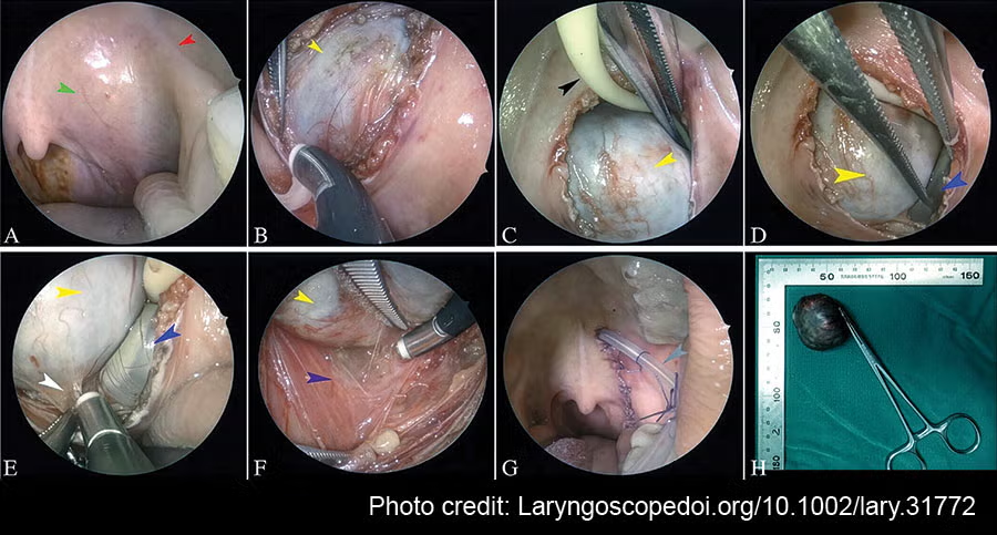
The endoscopic-assisted transoral PSBT resection protocol involved routine anesthesia, meticulous draping, and disinfection. A mouth gag was used to ensure optimal exposure of the surgical field. After irrigating the surgical area with diluted povidone-iodine solution and normal saline, an incision was made on the inner side of the pterygomandibular suture, at the most prominent part of the tumor. This incision was strategically made parallel to the pterygomandibular suture and slightly longer than the tumor’s dimensions. A high-definition 70° endoscope and a low-temperature plasma surgical system were then used for deep dissection, reaching the tumor surface. Bleeding vessels encountered during this phase were managed with bipolar coagulation.
Once the tumor surface was reached, the plasma surgical system was used to dissect the tumor from its capsule. The key innovation of the WSD technique was then implemented. A disposable catheter was carefully inserted into the space between the tumor capsule and the surrounding tissue. Water was then injected into the catheter, creating a water sac that gently distended the space around the tumor. This dilation facilitated blunt dissection, separating the tumor from its adjacent structures for one to two minutes. During this water sac maintenance period, surgeons utilized a plasma knife head or a long bipolar suction hook, guided by the endoscope, to further dissect the connective tissues surrounding the tumor. After the water sac was withdrawn, endoscopic visualization guided further dissection of the tissue at the site of water sac expansion. The tumor was then meticulously dissected and removed.
Following tumor extraction, the surgical cavity underwent thorough irrigation with diluted povidone-iodine solution and sterilized water to ensure complete removal of any residual tumor fragments and to achieve hemostasis. Negative pressure drainage was then placed in the surgical cavity, and the drainage tube was secured to the buccal mucosa. A gastric tube was maintained for one week post-surgery to facilitate feeding, and prophylactic antibiotics were administered. The drainage tube was removed when the daily drainage volume decreased to less than 10 mL. Post-operative MRI was performed five to seven days after surgery to evaluate the surgical site. Patients were discharged once significant swelling and hematoma formation were absent.
For the control group, the tumor was dissected and separated using fingers and cotton swabs for blunt dissection, followed by the same subsequent surgical steps as in the WSD group.
Statistical Analysis and Outcome Measures
Statistical analysis was performed using SPSS 20.0 software. Continuous variables were presented as mean ± standard deviation and compared using independent sample t-tests. Categorical variables were analyzed using the chi-square test. A p-value of less than 0.05 was considered statistically significant.
The primary outcome variables collected for analysis included:
- Operation time
- Intra-operative blood loss
- Drainage volume on the first post-operative day
- Total duration of drainage
- Total drainage volume
- Incidence of surgical complications
- Tumor recurrence rate
- Other prognostic data
Results: Tangible Benefits of the WSD Technique
The study’s findings revealed that all 32 patients successfully underwent tumor resection via the transoral approach, and post-operative pathology confirmed the absence of malignant lesions in all cases. The WSD group comprised 17 patients (53.1%), while the control group consisted of 15 patients (46.9%). A thorough comparison of baseline data, including gender, age, tumor size, and pathological type, showed no statistically significant differences between the two groups (all p > 0.05). This ensured that the observed differences in outcomes were attributable to the surgical technique rather than pre-existing patient characteristics.

The most striking results emerged when comparing the quantitative surgical outcomes between the WSD group and the control group. The WSD group demonstrated significantly lower values for operation time, intra-operative blood loss, drainage volume on the first post-operative day, total duration of drainage, and total drainage volume in the surgical area. All these differences were statistically significant (all p < 0.05). This indicates that the WSD technique facilitated a more efficient and less traumatic surgical dissection.
Crucially, the study reported no surgery-related complications in either group during the recovery period. Furthermore, at the six-month follow-up, MRI scans revealed no residual tumor or recurrence in any of the patients. Local surgery-related dysfunction was also absent during the follow-up period. Figure 2, illustrating typical pre-operative and post-operative MRI findings from a patient in the WSD group, visually confirms the successful tumor removal and the absence of complications.
Discussion: Enhancing Surgical Precision and Patient Recovery
The results of this study strongly suggest that the water sac dilation (WSD) method is a valuable adjunct to the endoscopy-assisted transoral approach for resecting benign parapharyngeal space tumors. The mechanism by which WSD enhances surgical outcomes appears to be multifaceted. By creating a controlled distension of the surgical space, the water sac gently separates the tumor from surrounding vital structures. This separation not only aids in blunt dissection, minimizing the need for aggressive cutting and reducing the risk of inadvertent injury to nerves and blood vessels, but also provides a clearer view of the tumor capsule and its boundaries. This enhanced visualization and gentle dissection likely contribute to reduced operative time and blood loss.
The significantly lower post-operative drainage volumes and shorter drainage durations in the WSD group are indicative of less tissue trauma and a more efficient closure of surgical planes. This, in turn, can lead to reduced post-operative swelling and discomfort, accelerating patient recovery and enabling earlier hospital discharge. The absence of surgery-related complications in both groups is a testament to the overall safety of the transoral approach for these tumors when performed by experienced teams, but the WSD technique appears to further refine this safety profile.
The success of the transoral approach, particularly with the WSD technique, aligns with the broader trend in surgical practice towards minimally invasive procedures. The avoidance of external scarring is a significant benefit for patients, improving their cosmetic outcomes and potentially reducing psychological distress. For PSBTs, which can occur in younger patients, cosmetic considerations are often paramount.
Implications and Future Directions
The findings of this study have significant clinical implications. The WSD method offers a practical and effective way to overcome some of the inherent challenges of endoscopic surgery in deep, confined spaces like the parapharyngeal space. The development of specialized WSD instruments tailored specifically for these procedures could further optimize surgical outcomes, potentially increasing the applicability of the transoral approach to a wider range of PSBTs.
While this study provides compelling evidence for the utility of WSD, further research with larger patient cohorts and longer follow-up periods would be beneficial to confirm these findings and explore its application in other challenging surgical sites. Investigating the cost-effectiveness of WSD compared to traditional methods would also be a valuable area for future research.
In conclusion, the integration of the water sac dilation method into the endoscopy-assisted transoral approach for benign parapharyngeal space tumor resection represents a significant advancement in surgical technique. It effectively minimizes intra-operative injury, enhances surgical efficiency, and expedites post-operative recovery, offering a less invasive and more patient-friendly alternative to traditional open surgeries. This innovation holds substantial promise for improving the management of these rare but surgically demanding tumors.