
The cerebellum, a region located at the base of the brain, is involved in nearly 80% of MS cases. Damage to this area manifests as some of the most debilitating symptoms of the disease, including tremors, unsteady gait, and a loss of fine motor skills. As healthy tissue within the cerebellum is gradually eroded, these symptoms often transition from intermittent "flares" to a state of permanent, progressive decline. The UCR study, led by Seema Tiwari-Woodruff, a professor of biomedical sciences, provides a new biological framework for understanding this transition, suggesting that the loss of myelin—the protective insulation around nerve fibers—triggers a catastrophic failure in the mitochondria of Purkinje cells.
The Neurological Energy Crisis: A New Frontier in MS Research
Multiple sclerosis is traditionally defined by the dual processes of inflammation and demyelination. In a healthy nervous system, the myelin sheath acts much like the plastic coating on an electrical wire, ensuring that nerve impulses travel rapidly and efficiently between the brain and the rest of the body. In MS, the immune system mistakenly attacks this sheath, leading to "short circuits" in neural communication. However, the UCR research highlights that demyelination is not merely a structural problem; it is an energetic one.
Mitochondria are the "powerhouses" of the cell, responsible for generating adenosine triphosphate (ATP), the chemical energy that fuels almost every biological process. When a nerve fiber loses its myelin insulation, it requires significantly more energy to transmit signals. Ironically, the UCR study found that at the very moment these neurons need more power, their mitochondria begin to fail. By analyzing the mitochondrial protein COXIV, researchers observed a significant decrease in mitochondrial activity within demyelinated Purkinje cells. This suggests that the cells are essentially "starving" of energy, leading to a programmed cell death that further cripples the patient’s motor functions.
Purkinje Neurons: The Fragile Architects of Movement
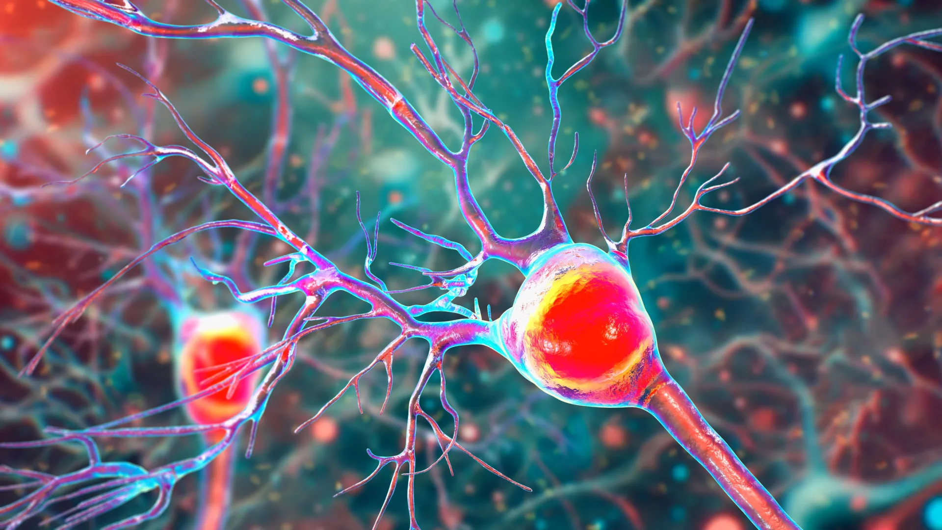
To understand the impact of this research, one must understand the unique role of Purkinje cells. These are among the largest and most complex neurons in the human brain, characterized by an extensive, tree-like thicket of branches known as dendrites. These branches allow a single Purkinje cell to receive and process an enormous amount of sensory information, which it then uses to coordinate smooth, precise movements—everything from the subconscious act of walking to the complex finger movements required to play a musical instrument.
"Inside the cerebellum are special cells called Purkinje neurons," Professor Tiwari-Woodruff explained. "These large, highly active cells help coordinate smooth, precise movements—like dancing, throwing a ball, or even just walking. They’re essential for balance and fine motor skills."
The UCR study found that in MS patients, these neurons undergo a tragic transformation before they eventually die. The researchers observed that the dendritic branches of Purkinje cells become stunted and sparse, a process that correlates with the loss of myelin. As these neurons lose their ability to process information and generate energy, they eventually perish. The resulting condition, known as ataxia, leaves patients struggling with coordination and stability, often requiring the use of mobility aids as the disease progresses.
Mapping Disease Progression Through Experimental Models
The research team utilized a multi-faceted approach to reach their conclusions, combining the study of human tissue with longitudinal observations in animal models. To track the progression of the disease in real-time, the team employed the experimental autoimmune encephalomyelitis (EAE) mouse model. This model mimics many of the pathological features of human MS, allowing scientists to observe the chronological stages of neurodegeneration that are difficult to capture in living human patients.
The timeline established by the EAE model revealed a distinct sequence of events. Demyelination and mitochondrial dysfunction appear early in the disease process, creating a period of cellular "distress" where the neurons are damaged but still alive. However, as the disease reaches its chronic phase, the cumulative lack of energy and the absence of myelin insulation lead to the widespread death of Purkinje cells. This finding is significant because it suggests a "window of opportunity" for medical intervention. If treatments can be developed to bolster mitochondrial health or protect myelin in the early stages, the eventual death of the brain cells might be prevented.
"The remaining neurons don’t work as well because their mitochondria start to fail," Tiwari-Woodruff noted. "These problems—less energy, loss of myelin, and damaged neurons—start early, but the actual death of the brain cells tends to happen later. The loss of energy in brain cells seems to be a key part of what causes damage in MS."
From Postmortem Analysis to Patient Care: The Research Methodology
A cornerstone of the study was the analysis of postmortem cerebellar tissue from individuals who had been diagnosed with secondary progressive MS, the stage of the disease where disability increases steadily. These samples, provided by the National Institutes of Health’s NeuroBioBank and the Cleveland Clinic, were compared against tissue from healthy donors of similar ages.
The histopathological analysis revealed a stark contrast. The MS-affected tissue showed clear evidence of chronic demyelination and a marked reduction in COXIV protein levels. By comparing these human findings with the data from the EAE mouse models, the researchers were able to confirm that the mitochondrial failure seen in mice was a faithful representation of the human disease state. This validation reinforces the EAE model’s value as a tool for testing future neuroprotective therapies.
The study was a collaborative effort, involving graduate student Kelley Atkinson, who conducted much of the primary research, alongside Shane Desfor, Micah Feria, Maria T. Sekyia, Marvellous Osunde, Sandhya Sriram, Saima Noori, Wendy Rincón, and Britany Belloa. The work was supported by funding from the National Multiple Sclerosis Society, highlighting the importance of non-profit investment in high-level academic research.
A Paradigm Shift in Treatment Strategies
Currently, most FDA-approved treatments for multiple sclerosis are "immunomodulatory," meaning they work by suppressing or altering the immune system to reduce the frequency of inflammatory relapses. While these drugs are effective at reducing new lesions in the brain and spinal cord, they have historically been less effective at stopping the "slow burn" of neurodegeneration that occurs in progressive MS.
The UCR findings suggest that a new class of treatments may be necessary—ones that focus on "neuroprotection" rather than just "immunosuppression." By targeting mitochondrial health, researchers hope to create therapies that can "supercharge" distressed neurons, giving them the energy required to survive the inflammatory environment of an MS brain.
"Our findings offer critical insights into the progression of cerebellar dysfunction in MS," Tiwari-Woodruff said. "Targeting mitochondrial health may represent a promising strategy to slow or prevent neurological decline and improve quality of life for people living with MS."
This shift in strategy could lead to the development of "mitochondrial cocktails"—supplements or medications designed to stabilize the COXIV protein or enhance ATP production. Furthermore, the research opens the door to therapies that promote "remyelination," the process of repairing the damaged myelin sheath, which would in turn reduce the metabolic strain on the Purkinje cells.
The Path Forward: Protecting White Matter and Support Cells
While the current study focused heavily on Purkinje cells, the UCR team is already looking toward the broader "ecosystem" of the brain. The cerebellum is not just composed of neurons; it also relies on support cells such as oligodendrocytes and astrocytes. Oligodendrocytes are the cells responsible for creating myelin, while astrocytes provide structural support and regulate the chemical environment of the brain.
The research team is now investigating whether mitochondrial damage extends to these support cells. If the "helper" cells of the brain are also suffering from energy failure, it creates a cycle of decay where the neurons cannot be repaired because the repair cells themselves are dying.
"To answer this, one of our ongoing research projects is focused on studying mitochondria in specific types of brain cells in the cerebellum," Tiwari-Woodruff said. "Such research can open the door to finding ways to protect the brain early on—like boosting energy in brain cells, helping them repair their protective myelin coating, or calming the immune system before too much damage is done."
The Socio-Economic Necessity of Sustained Research Funding
The implications of this research extend beyond the laboratory and into the realm of public policy and healthcare economics. MS is a disease that often strikes individuals in the prime of their lives, leading to decades of medical costs and lost productivity. By identifying a specific mechanism for disability—mitochondrial failure in the cerebellum—researchers are providing a roadmap to reduce the long-term burden of the disease.
However, Professor Tiwari-Woodruff warned that the pace of such breakthroughs is entirely dependent on consistent financial support for the sciences. In a climate of fluctuating federal budgets, she emphasized that medical progress is not a luxury but a necessity.
"Cutting funding to science only slows progress when we need it most," she stated. "Public support for research matters now more than ever."
As the scientific community digests the findings published in PNAS, the focus turns to how these insights can be translated into clinical trials. For the millions of people living with MS, the hope is that by understanding the "energy crisis" within their own brains, medicine can finally find a way to keep the lights on for the neurons that govern their movement, balance, and independence. The UCR study stands as a testament to the power of basic science to illuminate the path toward more effective, targeted treatments for one of neurology’s most enduring challenges.