

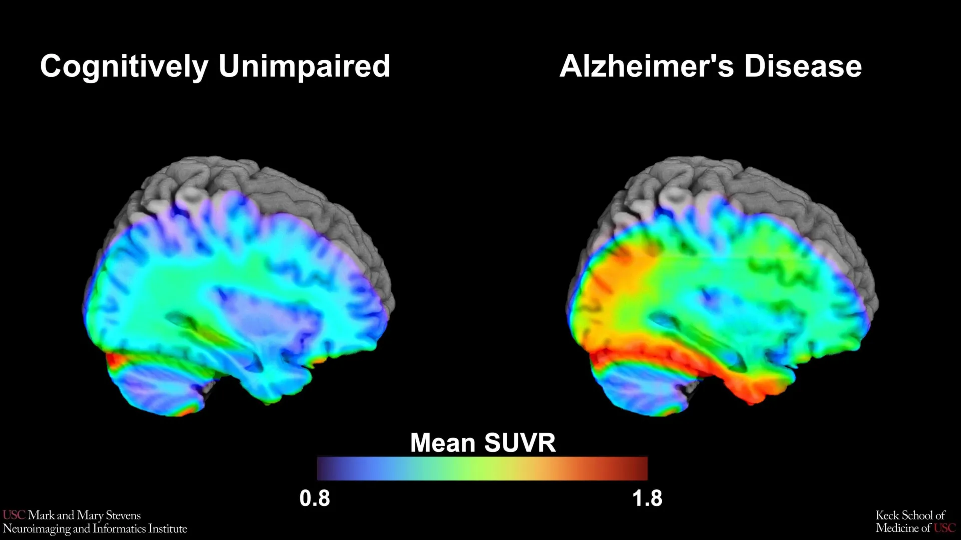
The study is a cornerstone of the Health and Aging Brain Study-Health Disparities (HABS-HD), a massive multi-university collaboration led by the University of North Texas Health Science Center and supported by the National Institute on Aging (NIA). By leveraging advanced Tau Positron Emission Tomography (PET) scans, the USC-led team analyzed a cohort of over 675 older adults to determine the precise threshold of protein accumulation that signals clinically relevant cognitive impairment.
The Biological Architecture of Alzheimer’s Disease
To understand the significance of the new benchmark, it is essential to distinguish between the two primary proteins associated with Alzheimer’s disease: amyloid-beta and tau. For decades, the "amyloid cascade hypothesis" dominated research, suggesting that the buildup of amyloid plaques was the primary driver of the disease. However, recent clinical developments have shifted focus toward tau proteins. While amyloid plaques often appear years before symptoms manifest, tau "tangles"—which form inside neurons—are more closely correlated with the actual onset of memory loss and cognitive decline.
The USC study utilized a next-generation imaging tracer known as 18F-PI-2620. This radioactive agent is designed to bind specifically to tau deposits in the brain, allowing them to glow during a PET scan. By quantifying this glow, researchers can measure the density of tau in specific regions, most notably the medial temporal lobe. The medial temporal lobe is a critical area for memory processing and is typically one of the first regions to show tau accumulation in the early stages of Alzheimer’s.
Establishing the Tau Cut-Point: A New Diagnostic Metric
The primary objective of the research was to establish a "cut-point"—a numerical threshold of tau protein density that separates "normal" aging from "pathological" Alzheimer’s progression. Establishing such a metric is vital for both clinical trials and future bedside diagnostics. Without a standardized cut-point, clinicians struggle to determine whether a patient’s tau levels are high enough to warrant aggressive intervention or inclusion in experimental drug trials.
In their analysis of 675 participants, the research team compared the tau PET scans of individuals classified as cognitively impaired with those who tested as cognitively normal through standardized neuropsychological assessments. The data revealed a specific threshold in the medial temporal lobe that strongly indicated the presence of Alzheimer’s-related impairment.
However, the efficacy of this cut-point was found to be contingent upon the presence of another factor: amyloid-beta. The tau benchmark was highly accurate in identifying cognitive impairment only when the participant also tested positive for amyloid plaques. This "dual-protein" requirement reinforces the current medical understanding that Alzheimer’s is a multi-faceted disease where different proteins interact to drive neurodegeneration.
Racial and Ethnic Disparities in Biomarker Reliability
One of the most striking findings of the study involves the divergence in results across different demographic groups. While the tau cut-point was a reliable indicator for Hispanic and non-Hispanic White participants, it failed to perform with the same accuracy in the non-Hispanic Black cohort.
"In non-Hispanic Black participants, the tau cut-point did not perform as expected," noted senior author Meredith N. Braskie, PhD, an assistant professor of neurology at USC. "This suggests that other pathologies or conditions may be driving cognitive decline in this group."
This finding is of paramount importance because Black Americans are statistically twice as likely to develop Alzheimer’s or other dementias as White Americans, yet they have historically been underrepresented in clinical research. The failure of the tau benchmark in this group suggests that the biological "signature" of cognitive decline in Black populations may involve a different balance of vascular issues, inflammation, or other proteinopathies that are not yet fully understood.
Lead author Victoria R. Tennant, a PhD candidate in USC’s Neuroscience Graduate Program, emphasized that these findings highlight the limitations of a "one-size-fits-all" approach to neurology. "The limited reliability of tau as an indicator of cognitive impairment in non-Hispanic Black participants highlights the need for more diverse populations in research and for future studies to examine both biological and social determinants of Alzheimer’s disease," Tennant stated.
Chronology and Context of the HABS-HD Initiative
The Health and Aging Brain Study-Health Disparities (HABS-HD) was launched to address the historical lack of diversity in Alzheimer’s research. Most existing data on AD biomarkers were derived from cohorts that were over 90% non-Hispanic White. HABS-HD was designed from the ground up to be the most comprehensive study of AD in a diverse community, specifically recruiting large numbers of Hispanic and Black participants.
The timeline of this research reflects a broader shift in the National Institutes of Health (NIH) strategy. Over the last decade, the NIA has significantly increased funding for health disparity research, recognizing that treatments developed for one demographic may not be effective for all. The USC study represents a major milestone in this multi-year effort, moving from general data collection to the establishment of specific, actionable diagnostic benchmarks.
The study’s reliance on the 18F-PI-2620 tracer also represents a technological evolution. Earlier tau tracers often suffered from "off-target binding," where the tracer would stick to areas of the brain unrelated to Alzheimer’s, creating "noise" in the data. The 18F-PI-2620 tracer is part of a newer generation of imaging tools that provide much clearer, more specific maps of tau distribution, allowing for the precise calculations required to set a cut-point.
Implications for Future Clinical Trials and Treatment
The establishment of a tau cut-point has immediate implications for the pharmaceutical industry. Currently, several high-profile Alzheimer’s drugs, such as lecanemab (Leqembi) and donanemab, focus on clearing amyloid from the brain. However, the next frontier of treatment involves "tau-targeting" therapies.
For these new drugs to be tested effectively, researchers must be able to identify trial participants who have a specific, measurable amount of tau in their brains. If a trial includes people with too little tau, the drug’s effects may not be visible; if it includes people with too much, the damage may already be irreversible. The USC team’s benchmark provides a "Goldilocks" zone for recruitment, ensuring that the right patients are selected for the right trials.
Furthermore, the study’s findings regarding the role of amyloid suggest that future treatments may need to be "combination therapies." If tau’s impact on cognition is most pronounced in the presence of amyloid, then clearing both proteins simultaneously might be the most effective path forward.
Analyzing the Role of Social Determinants
The discrepancy in how tau markers function in Black participants has led the research team to call for a broader investigation into the "social determinants of health." Biological markers do not exist in a vacuum; they are influenced by a lifetime of environmental factors, including access to healthcare, diet, chronic stress, and exposure to pollutants.
Arthur W. Toga, PhD, director of the Stevens INI, highlighted that HABS-HD is uniquely positioned to explore these intersections. "HABS-HD has already produced key findings related to ethnic variations in AD biomarkers, the influences of social determinants on cognitive health, and vascular contributions to dementia," Toga said. "We hope this work will lead to more personalized care and better outcomes for all communities."
For instance, higher rates of hypertension and diabetes in certain communities can lead to "vascular dementia," which may coexist with or mimic the symptoms of Alzheimer’s. If a Black patient’s cognitive decline is being driven primarily by vascular damage rather than tau tangles, a tau-focused treatment would be ineffective. This study provides the empirical evidence needed to shift clinical focus toward these multi-pathology models.
A Collaborative Effort in Neuroimaging
The study was a massive undertaking involving dozens of experts from across the United States. In addition to the USC team, contributors included researchers from the University of North Texas Health Science Center, the University of Wisconsin-Madison, Washington University in St. Louis, and the University of California, San Francisco.
The research was supported by multiple grants from the National Institute on Aging and the Office of the Director at the NIH. This level of federal support underscores the urgency of the Alzheimer’s crisis, which currently affects more than 6 million Americans—a number projected to rise to nearly 13 million by 2050 without significant medical breakthroughs.
By refining how we see the brain through tau PET imaging, the researchers at USC and their partners are not just identifying a disease; they are mapping the nuances of human diversity within the most complex organ in the body. The establishment of this tau cut-point marks a transition from a generalized understanding of Alzheimer’s to a more precise, inclusive, and personalized era of neurodegenerative medicine. As researchers continue to untangle the relationship between tau, amyloid, and ethnicity, the goal remains clear: ensuring that the future of Alzheimer’s care is effective for every patient, regardless of their background.