
The Critical Role of Purkinje Cells in Neurological Health
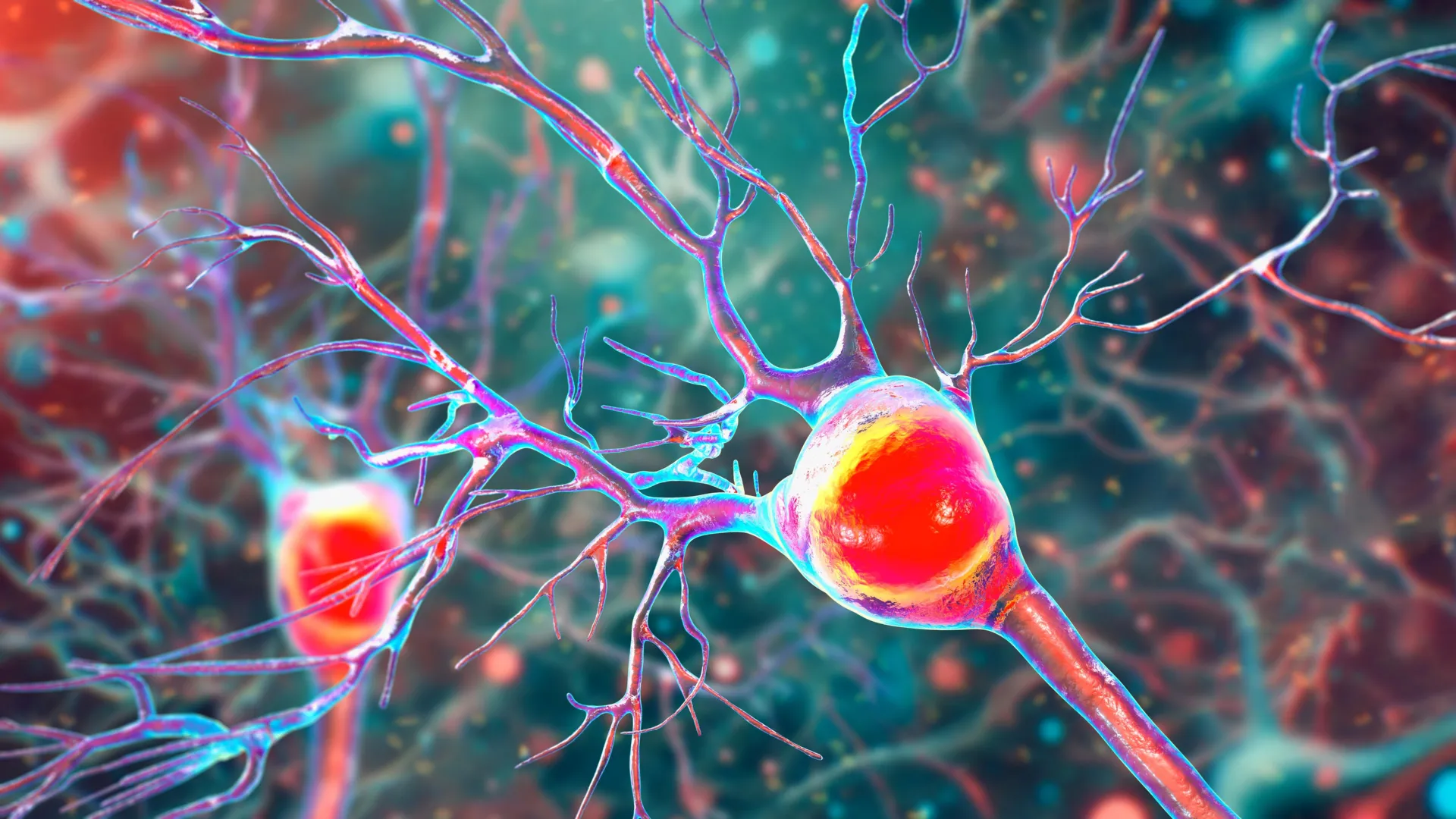
To understand the impact of the UC Riverside findings, it is necessary to recognize the specialized function of Purkinje cells. These are among the largest and most complex neurons in the human brain, characterized by an intricate, tree-like branching structure called a dendritic arbor. This structure allows a single Purkinje cell to receive and process an immense amount of information from other neurons, making them the primary output cells of the cerebellar cortex.
"Inside the cerebellum are special cells called Purkinje neurons," explained Professor Tiwari-Woodruff. "These large, highly active cells help coordinate smooth, precise movements—like dancing, throwing a ball, or even just walking. They’re essential for balance and fine motor skills." Because these cells are so metabolically active, they require a constant and robust supply of energy. When that energy supply is interrupted, the cells cannot maintain their complex structures or perform their signaling duties, eventually leading to cell death. In MS patients, the gradual disappearance of these neurons is a primary driver of permanent disability.
Demyelination and the Cascade of Mitochondrial Failure
The hallmarks of MS are inflammation and demyelination. Myelin is a fatty, insulating substance that wraps around nerve fibers (axons), much like the plastic coating on an electrical wire. This insulation is vital for the rapid and efficient transmission of electrical impulses. In MS, the immune system mistakenly attacks this myelin, leading to "short circuits" in the brain’s communication network.
However, the research conducted by Tiwari-Woodruff’s team, including lead graduate student Kelley Atkinson, suggests that the damage goes deeper than the surface insulation. The study proposes that the inflammatory environment and the loss of myelin disrupt the internal machinery of the Purkinje cells—specifically the mitochondria. By analyzing postmortem cerebellar tissue from individuals who had secondary progressive MS (SPMS), the researchers discovered a significant deficiency in a mitochondrial protein known as COXIV (Cytochrome c oxidase subunit 4).
COXIV is essential for the electron transport chain, the process by which mitochondria convert nutrients into adenosine triphosphate (ATP), the primary energy currency of the cell. The study found that in areas where myelin was lost, Purkinje cells showed a marked decrease in COXIV expression. This "energy failure" correlates directly with the thinning of the cells’ dendritic branches and their eventual death.
Evidence from the EAE Mouse Model
To validate their findings from human tissue, the researchers utilized the Experimental Autoimmune Encephalomyelitis (EAE) mouse model. EAE is a widely accepted surrogate for studying MS because it replicates the inflammatory and demyelinating aspects of the human disease. By tracking the progression of EAE in mice, the team was able to establish a clear chronology of how cerebellar damage unfolds.
The observations in the mouse model mirrored the human data with striking accuracy. Early in the disease process, the mice exhibited demyelination in the cerebellum. This was followed closely by a decline in mitochondrial function within the Purkinje cells. As the disease transitioned into a chronic phase, the researchers observed a steady decline in the number of Purkinje cells, which coincided with the onset of severe movement problems in the mice.
"The remaining neurons don’t work as well because their mitochondria, the energy-producing parts, start to fail," Tiwari-Woodruff noted. "We also saw that the myelin breaks down early in the disease. These problems—less energy, loss of myelin, and damaged neurons—start early, but the actual death of the brain cells tends to happen later, as the disease becomes more severe." This timeline is crucial because it suggests a "window of opportunity" where therapeutic intervention could potentially save neurons before they are lost forever.
Supporting Data and the Global Impact of MS
The implications of this research are significant when viewed against the backdrop of global health statistics. MS is the leading cause of non-traumatic disability in young adults, with most patients diagnosed between the ages of 20 and 40. While many patients initially experience relapsing-remitting MS (RRMS), characterized by periods of symptoms followed by recovery, many eventually transition to secondary progressive MS, where disability worsens steadily without periods of remission.
According to the National Multiple Sclerosis Society, which funded the UC Riverside study, the economic burden of MS in the United States alone exceeds $85 billion annually, including direct medical costs and lost productivity. The cerebellar symptoms—ataxia and tremors—are among the most difficult to treat and have a profound impact on a patient’s quality of life and independence. By identifying mitochondrial failure as a driver of Purkinje cell loss, the research provides a specific biological target for drugs that could stabilize the disease in its progressive stages.
Implications for Future Therapeutic Strategies
Currently, most MS treatments are immunomodulatory, meaning they focus on reducing the frequency and severity of immune system attacks. While these are effective for RRMS, they have shown limited success in preventing the long-term neurodegeneration seen in progressive MS. The UC Riverside study suggests that a "neuroprotective" approach—one that focuses on the health and energy production of the neurons themselves—is necessary.
"Targeting mitochondrial health may represent a promising strategy to slow or prevent neurological decline and improve quality of life for people living with MS," said Tiwari-Woodruff. This could involve the development of compounds that boost COXIV activity, antioxidants that protect mitochondria from oxidative stress caused by inflammation, or therapies that promote the repair of myelin (remyelination) to restore the metabolic environment of the neuron.
Furthermore, the research team is expanding its scope to determine if mitochondrial failure is a universal problem across different brain cell types. They are currently investigating whether similar energy deficits occur in oligodendrocytes (the cells responsible for creating myelin) and astrocytes (cells that support the blood-brain barrier and provide nutrients to neurons). If mitochondrial dysfunction is widespread across these cell types, it would suggest that MS is as much a metabolic "energy crisis" in the brain as it is an autoimmune disorder.
The Necessity of Sustained Scientific Investment
The study concludes with a call to action regarding the broader scientific landscape. Professor Tiwari-Woodruff emphasized that the complexity of MS requires long-term, sustained research efforts. The use of postmortem tissue, provided by the National Institutes of Health’s NeuroBioBank and the Cleveland Clinic, highlights the importance of patient participation and infrastructure in medical breakthroughs.
"Cutting funding to science only slows progress when we need it most," Tiwari-Woodruff stated, addressing the ongoing challenges of securing research grants. "Public support for research matters now more than ever."
As the scientific community moves forward, the focus will likely shift toward "early-intervention" strategies. By understanding that mitochondrial failure begins long before cell death occurs, clinicians may one day be able to use advanced imaging or biomarkers to identify patients at risk of cerebellar decline. Intervening at this stage—by boosting cellular energy or calming the local immune response—could potentially allow patients to maintain their mobility and balance for decades longer than is currently possible.
This research, authored by Tiwari-Woodruff and Atkinson alongside a collaborative team including Shane Desfor, Micah Feria, and others, represents a significant step toward a future where MS is no longer a sentence of progressive physical decline, but a manageable condition where the brain’s own "powerhouses" are protected and preserved.