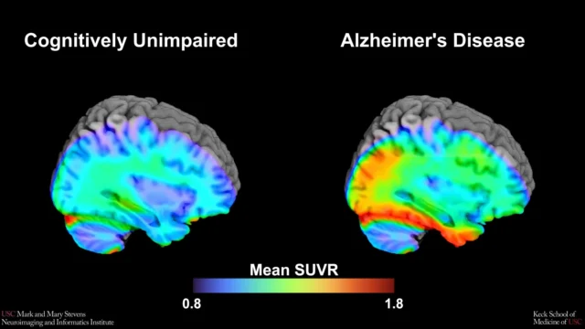
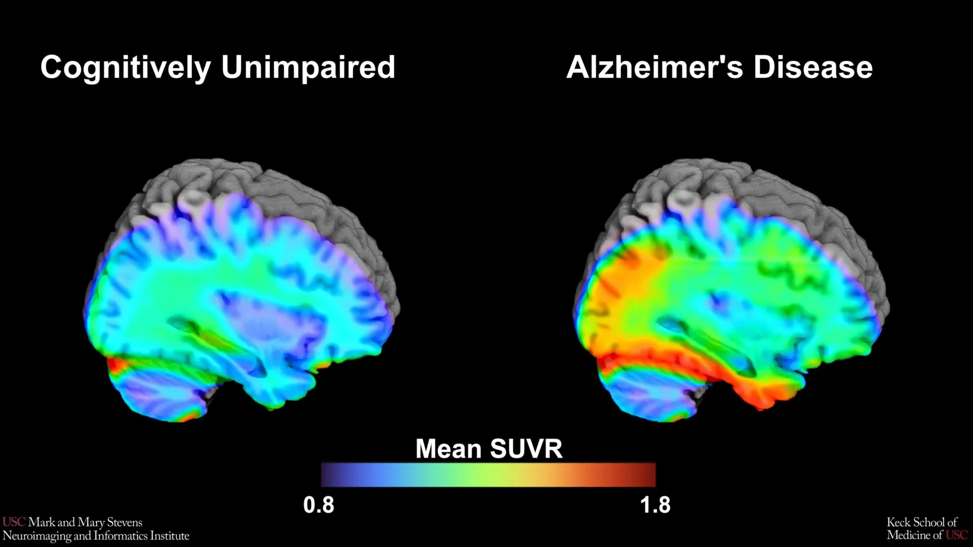
The study centers on the utilization of tau positron emission tomography (PET) scans, a sophisticated imaging modality that allows scientists to observe the accumulation of specific proteins within the living brain. For decades, Alzheimer’s research focused primarily on amyloid-beta plaques. However, modern neuroscience has increasingly recognized that tau protein "tangles" are more closely correlated with the actual onset of cognitive symptoms and the death of neurons. By establishing a "cut-point"—a specific threshold of tau protein density—the researchers aimed to create a binary diagnostic tool that could reliably separate healthy aging from the early pathological stages of Alzheimer’s disease (AD).
The Biological Significance of Tau and the PET Imaging Evolution
Alzheimer’s disease is characterized by the progressive accumulation of two abnormal protein structures: amyloid-beta plaques, which gather between neurons, and tau tangles, which form inside the neurons themselves. While amyloid is often considered one of the earliest signs of the disease, its presence does not always guarantee immediate cognitive decline. Tau, conversely, is viewed as the "executioner" protein; its spread through the brain’s cortex mirrors the progression of memory loss and functional impairment.
To visualize this process, the USC team employed an advanced radioactive tracer known as 18F-PI-2620. This tracer is designed to bind specifically to tau aggregates, allowing them to glow on a PET scan. By measuring the "standardized uptake value ratio" (SUVR), researchers can quantify the density of tau in specific brain regions. The Stevens INI study focused heavily on the medial temporal lobe, a region deep within the brain that includes the hippocampus and is responsible for memory formation. This area is typically the first to show tau accumulation in the early stages of Alzheimer’s.
The establishment of a "cut-point" is a foundational step in transforming raw imaging data into a clinical tool. In medical diagnostics, a cut-point acts as a line in the sand; individuals whose protein levels fall above the line are considered "tau positive," indicating a high likelihood of Alzheimer’s pathology, while those below are "tau negative." Prior to this study, many such benchmarks were derived from small, homogenous groups, often failing to account for the biological and environmental diversity of the broader population.
Study Methodology and Participant Demographics
The research utilized data from 675 older adults enrolled in the HABS-HD study, a multi-university collaboration led by the University of North Texas Health Science Center. HABS-HD is specifically designed to investigate the impact of health disparities on Alzheimer’s and related dementias. The cohort included a robust representation of Hispanic, non-Hispanic White, and non-Hispanic Black participants, making it one of the most demographically inclusive neuroimaging studies to date.
The methodology involved a rigorous comparison between two primary groups: those who were cognitively impaired (demonstrating measurable deficits in memory, language, or executive function) and those who were cognitively unimpaired. By analyzing the tau PET scans of these individuals, the researchers sought to find the exact level of tau in the medial temporal lobe that best predicted whether an individual would fall into the "impaired" category.
A critical layer of the study involved the presence of amyloid. The researchers found that the predictive power of the tau cut-point was significantly enhanced when amyloid-beta was also present in the brain. This reinforces the "amyloid cascade hypothesis," which suggests that amyloid may act as a catalyst that triggers or accelerates the spread of tau, leading to the symptomatic stage of the disease.
Findings: Successes in Hispanic and White Populations
The results of the study provided a clear benchmark for two of the three groups studied. For Hispanic and non-Hispanic White participants, the identified tau cut-point in the medial temporal lobe proved to be a highly effective indicator of cognitive impairment, provided that amyloid was also present. This finding is particularly significant for the Hispanic community, which has historically been underrepresented in Alzheimer’s research despite being at a higher risk for the disease compared to non-Hispanic Whites.
"Our tau cut-point was able to distinguish whether study participants had cognitive impairment—but only when another abnormal protein, amyloid, was also present in those with cognitive impairment, and only in Hispanic and non-Hispanic White participants," explained senior author Meredith N. Braskie, PhD, assistant professor of neurology at the Keck School of Medicine of USC.
The success of the 18F-PI-2620 tracer in these groups suggests that the medial temporal lobe remains a "ground zero" for AD pathology across these populations. Establishing this benchmark allows clinicians to move toward more standardized diagnostic criteria, potentially leading to earlier interventions and more accurate recruitment for clinical trials targeting tau protein.
The Diagnostic Gap: Non-Hispanic Black Participants
While the study achieved its goals for Hispanic and White populations, it revealed a troubling discrepancy regarding non-Hispanic Black participants. In this group, the established tau cut-point did not reliably distinguish between those with and without cognitive impairment. Even when tau levels were elevated, they did not correlate with memory loss and cognitive decline in the same way observed in other groups.
This finding suggests that the biological drivers of dementia in Black populations may be more complex than the traditional amyloid-tau model. "In non-Hispanic Black participants, the tau cut-point did not perform as expected," Dr. Braskie noted. "This suggests that other pathologies or conditions may be driving cognitive decline in this group."
Researchers point toward several potential factors for this divergence, including vascular contributions to dementia (such as hypertension and small vessel disease), chronic inflammation, and the impact of lifelong social stressors. These "social determinants of health" can lead to "weathering"—a process where chronic stress causes premature biological aging. If cognitive decline in Black participants is being driven by vascular damage or other non-Alzheimer’s pathologies, then a diagnostic tool focused solely on tau protein will inevitably fall short.
Official Reactions and the Path Toward Health Equity
The implications of this research extend far beyond the laboratory. By highlighting where current diagnostic models fail, the USC team is calling for a paradigm shift in how Alzheimer’s is studied and treated.
Lead author Victoria R. Tennant, a PhD candidate in USC’s Neuroscience Graduate Program, emphasized the dual nature of the study’s results. "While our findings support prior research linking medial temporal lobe tau to cognitive impairment, establishing a cut-point in this region using 18F-PI-2620 marks an important step toward defining tau positivity for both research and clinical applications," Tennant stated. "At the same time, the limited reliability of tau as an indicator of cognitive impairment in non-Hispanic Black participants highlights the need for more diverse populations in research and for future studies to examine both biological and social determinants of Alzheimer’s disease."
Arthur W. Toga, PhD, director of the Stevens INI, underscored the importance of the HABS-HD framework in achieving these insights. "This type of imaging is critical for understanding who is at risk and how the disease develops," Toga said. "These findings are just the latest to come from HABS-HD, which is the most comprehensive study of Alzheimer’s disease and related dementias in diverse communities." Toga further noted that the project has already shed light on ethnic variations in biomarkers and vascular contributions to dementia, with the ultimate goal of fostering "personalized care and better outcomes for all communities."
Timeline and Institutional Support
The HABS-HD study is an ongoing multi-year initiative that represents a massive logistical and financial commitment to public health. Supported by the National Institute on Aging (NIA), the study involves a network of institutions including the University of North Texas Health Science Center, the University of Wisconsin-Madison, Washington University in St. Louis, and the University of California, San Francisco.
The publication of these findings in Imaging Neuroscience marks a critical milestone in the study’s timeline, providing the first validated cut-points for the 18F-PI-2620 tracer in a diverse cohort. The research was made possible through several grants from the National Institutes of Health (NIH), totaling millions of dollars in federal investment aimed at solving the Alzheimer’s crisis.
Broader Impact and Future Implications
The discovery of a tau cut-point that works for Hispanic and White populations provides a new tool for pharmaceutical companies. As new "anti-tau" drugs enter clinical trials, having a reliable way to screen participants is essential. If researchers can identify "tau-positive" individuals before they show severe symptoms, they can test treatments that might stop the "fire" of tau from spreading through the brain.
However, the "failure" of the benchmark in Black populations is equally important for the future of medicine. It serves as a warning that a "one-size-fits-all" approach to Alzheimer’s diagnosis could lead to misdiagnosis or the exclusion of Black patients from relevant clinical trials. Future research must now pivot to identify the specific biomarkers—whether vascular, inflammatory, or genetic—that accurately predict cognitive decline in Black communities.
Furthermore, this study highlights the necessity of "multi-modal" diagnostics. Relying on a single protein (like tau) may not be enough. The future of Alzheimer’s care likely lies in a combination of PET scans, blood-based biomarkers, and an assessment of a patient’s vascular health and social history.
As the global population ages, the burden of Alzheimer’s disease is expected to triple by 2050. The work being done at the Keck School of Medicine of USC and through the HABS-HD collaboration ensures that the tools developed to fight this epidemic are effective for everyone, regardless of their ethnic or racial background. By refining these benchmarks, scientists are moving closer to a day when Alzheimer’s can be detected, categorized, and treated with the precision that the 21st century demands.