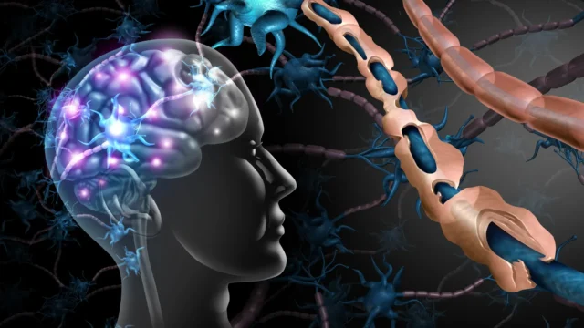
The study, led by Seema Tiwari-Woodruff, a professor of biomedical sciences at the UC Riverside School of Medicine, focuses on the cerebellum—a region at the base of the brain that serves as the command center for motor control, balance, and coordination. In approximately 80% of MS cases, the cerebellum becomes a site of intense inflammation and tissue loss. This damage manifests clinically as tremors, an unsteady gait, and difficulty with fine motor tasks, symptoms that often define the transition from relapsing-remitting MS to the more severe progressive stages of the disease.
The Role of Purkinje Cells in Motor Function
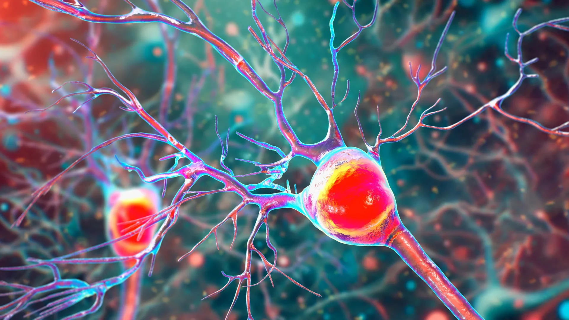
At the heart of the UCR study are Purkinje cells, some of the largest and most complex neurons in the human brain. Located within the cerebellar cortex, these cells are essential for the inhibitory control of motor movements. They act as a sophisticated "braking system," ensuring that muscle contractions are smooth, timed correctly, and proportional to the task at hand—whether that involves walking across a room or threading a needle.
"Inside the cerebellum are special cells called Purkinje neurons," Tiwari-Woodruff explained. "These large, highly active cells help coordinate smooth, precise movements—like dancing, throwing a ball, or even just walking. They’re essential for balance and fine motor skills."
Because Purkinje cells are so large and electrically active, they possess an extraordinarily high demand for energy. This makes them uniquely vulnerable to any disruption in their power supply. In the context of MS, the study found that as inflammation and demyelination take hold in the cerebellum, these neurons begin to wither and eventually die. This loss is the direct precursor to ataxia, a neurological sign characterized by a lack of voluntary coordination of muscle movements.
A Bioenergetic Crisis: The Mitochondrial Link
For decades, MS research focused primarily on the immune-mediated destruction of myelin, the fatty insulation that allows electrical signals to jump quickly from one node of a nerve to the next. However, the UCR team, spearheaded by graduate student Kelley Atkinson, shifted the lens toward the "powerhouses" of the cell: the mitochondria.
Mitochondria are responsible for producing adenosine triphosphate (ATP), the chemical energy that fuels almost every cellular process. The researchers discovered that in the demyelinated regions of the cerebellum, there is a significant drop in the levels of a vital mitochondrial protein known as COXIV (Cytochrome c oxidase subunit 4). This protein is a key component of the electron transport chain, the machinery that generates ATP.
When COXIV levels plummet, the Purkinje cells enter a state of energy failure. Without sufficient ATP, the cells cannot maintain their complex branching structures (dendrites), nor can they survive the toxic environment created by chronic inflammation. The UCR study proposes that this mitochondrial impairment is not merely a side effect of MS but a direct driver of cell death.
"Our study proposes that inflammation and demyelination in the cerebellum disrupt mitochondrial function, contributing to nerve damage and Purkinje cell loss," said Tiwari-Woodruff. "We observed a significant loss of the mitochondrial protein COXIV in demyelinated Purkinje cells, suggesting that mitochondrial impairment contributes directly to cell death and cerebellar damage."
Evidence from Human Tissue and Animal Models
To reach these conclusions, the research team employed a dual-track methodology, analyzing both human postmortem tissue and an established animal model of the disease.
The human component of the study involved the examination of cerebellar tissue from individuals who had been diagnosed with secondary progressive MS. These samples, provided by the National Institutes of Health’s NeuroBioBank and the Cleveland Clinic, allowed the researchers to observe the end-stage reality of the disease. They found that neurons in these patients had fewer branches and significant mitochondrial deficits compared to healthy control samples.
To understand how these changes develop over time, the team used the Experimental Autoimmune Encephalomyelitis (EAE) mouse model. EAE mimics many of the pathological features of human MS, including the immune system’s attack on the central nervous system. By tracking the mice through various stages of the disease, the researchers were able to establish a clear chronology of decline.
The data showed that myelin breakdown occurs early in the disease process. Shortly thereafter, mitochondrial function begins to fail. While the neurons initially survive these insults, they do so in a compromised state. The actual death of the Purkinje cells tends to occur later, as the disease enters a chronic, severe phase. This suggests a "window of opportunity" where intervention might be possible before the damage becomes irreversible.
"The remaining neurons don’t work as well because their mitochondria start to fail," Tiwari-Woodruff noted. "These problems—less energy, loss of myelin, and damaged neurons—start early, but the actual death of the brain cells tends to happen later. The loss of energy in brain cells seems to be a key part of what causes damage in MS."
Data and Statistical Context
The implications of this research are underscored by the broader epidemiological data regarding Multiple Sclerosis. MS is most commonly diagnosed in adults between the ages of 20 and 50, and it is significantly more prevalent in women than in men, with a ratio of approximately 3:1.
While life expectancy for MS patients has improved significantly over the last 50 years, the quality of life remains a major concern, particularly for those with progressive forms of the disease. Progressive MS accounts for about 10% to 15% of diagnoses at onset (Primary Progressive MS) and eventually affects a large portion of those initially diagnosed with Relapsing-Remitting MS (Secondary Progressive MS).
The economic impact is also substantial. In the United States alone, the total economic burden of MS—including direct medical costs and indirect costs like lost productivity—is estimated to be over $85 billion annually. Research that identifies specific pathways for preventing neuronal loss, such as the UCR mitochondrial study, is vital for reducing both the human and economic costs of the disease.
Implications for Future Therapeutics
The discovery that mitochondrial failure precedes cell death opens a new frontier for MS treatment. Current FDA-approved disease-modifying therapies (DMTs) are largely focused on modulating the immune system to reduce the frequency of relapses. While effective at slowing the early stages of MS, these treatments have historically been less successful at halting the steady neurodegeneration seen in progressive MS.
The UCR findings suggest that a multi-pronged approach may be necessary. "Targeting mitochondrial health may represent a promising strategy to slow or prevent neurological decline and improve quality of life for people living with MS," Tiwari-Woodruff stated.
Potential future treatments could include:
- Mitochondrial Boosters: Drugs designed to enhance the efficiency of the electron transport chain or increase the production of ATP in stressed neurons.
- Neuroprotective Agents: Compounds that shield Purkinje cells from the oxidative stress that results when mitochondria malfunction.
- Remyelination Therapies: While the study highlights mitochondria, the loss of myelin is still the initial trigger. Combining myelin repair with energy support could provide a synergistic effect.
Expanding the Research Horizon
The UCR team is not stopping at Purkinje cells. Their ongoing projects are looking at how mitochondrial damage affects other vital components of the cerebellum, such as oligodendrocytes (the cells that produce myelin) and astrocytes (star-shaped cells that provide structural and metabolic support to neurons).
"To answer this, one of our ongoing research projects is focused on studying mitochondria in specific types of brain cells in the cerebellum," Tiwari-Woodruff said. "Such research can open the door to finding ways to protect the brain early on—like boosting energy in brain cells, helping them repair their protective myelin coating, or calming the immune system before too much damage is done."
This holistic view of the "cerebellar environment" is crucial because MS is not just a disease of one cell type, but a failure of a complex biological ecosystem. By understanding how different cells interact and fail together, researchers hope to develop "cocktail" therapies that address inflammation, demyelination, and metabolic failure simultaneously.
The Vital Role of Research Funding
The study was supported by the National Multiple Sclerosis Society, a reminder of the critical role that non-profit and public funding play in medical breakthroughs. Tiwari-Woodruff took the opportunity to advocate for continued investment in the sciences, noting that the path from laboratory discovery to clinical treatment is long and requires sustained support.
"Cutting funding to science only slows progress when we need it most," she said. "Public support for research matters now more than ever."
As the scientific community digests the findings of the UCR study, the focus shifts toward translating these metabolic insights into tangible help for patients. For the 2.3 million people living with MS, the shift from viewing the disease as purely an immune problem to an energetic one offers a new beacon of hope. If the "powerhouses" of the brain can be kept running, the devastating loss of coordination and independence that characterizes progressive MS might one day be a thing of the past.
The research team involved in this significant work included Seema Tiwari-Woodruff and Kelley Atkinson, along with Shane Desfor, Micah Feria, Maria T. Sekyia, Marvellous Osunde, Sandhya Sriram, Saima Noori, Wendy Rincón, and Britany Belloa. Their collaborative effort represents a major step forward in the quest to demystify and eventually defeat Multiple Sclerosis.