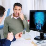

The Clinical Imperative for AI in Cholesteatoma Diagnosis
Cholesteatoma, a non-cancerous but invasive skin growth in the middle ear, arises from the retraction or migration of the tympanic membrane or external ear canal skin. Its insidious nature means early detection is paramount to prevent its destructive progression. The disease erodes the delicate ossicles, mastoid bone, and can even extend into adjacent structures like the facial nerve or brain, leading to serious sequelae such as facial paralysis, meningitis, and brain abscesses. Traditional diagnostic methods rely on a combination of clinical examination, audiometry, and imaging studies, primarily CT scans. However, the subtle differences in bony destruction and soft tissue density between cholesteatoma and inflammatory granulations or cholesteatoma mimics can lead to diagnostic ambiguity, impacting timely and appropriate treatment.
The advent of advanced AI techniques, specifically deep learning and convolutional neural networks (CNNs), has opened new avenues for image analysis. These algorithms can be trained on vast datasets of medical images to identify intricate patterns and features that might be imperceptible to the human eye. For cholesteatoma, this translates to the potential for automated detection, precise localization, and even staging of the disease, thereby aiding surgeons in pre-operative planning and potentially improving patient outcomes.
A Comprehensive Review of Existing Evidence
A systematic review, meticulously conducted according to the Preferred Reporting Items for Systematic Reviews and Meta-Analyses (PRISMA) guidelines, has synthesized the current evidence on AI’s efficacy in diagnosing cholesteatoma. The review, which searched major scientific databases including PubMed, Scopus, and Web of Science up to September 2025, identified seven studies that met stringent inclusion criteria. These studies investigated the application of AI, machine learning, or deep learning models for cholesteatoma diagnosis utilizing CT, magnetic resonance imaging (MRI), or otoscopic imaging. The research spanned multinational efforts, with contributing institutions located in Turkey, Japan, China, Morocco, and the United States, underscoring the global interest and collaborative efforts in this domain.
Key Findings: High Accuracy, Promising Architectures
The analysis revealed that AI-based models have demonstrated remarkable diagnostic accuracy for cholesteatoma detection and staging. A significant majority of the included studies employed sophisticated CNN architectures, a type of deep learning algorithm particularly adept at processing image data. Popular architectures cited in the review include DenseNet, MobileNetV2, ResNet50, Inception-V3, and Xception. These models were trained and validated on datasets to identify cholesteatoma from various imaging modalities.
CT Imaging: The Leading Modality
Temporal bone CT imaging emerged as the primary imaging modality utilized in the AI-driven cholesteatoma diagnosis research. Six of the seven studies focused on CT scans, leveraging their ability to provide detailed anatomical information. The internal validation performance of these CT-based AI models was generally exceptional, with accuracies often exceeding 90%. This high internal validation suggests that within the specific datasets used for training and testing, the AI models performed with impressive precision.
Specific examples of high performance were noted:
- ResNet50 achieved an accuracy of 93.3% in distinguishing between chronic otitis media with and without cholesteatoma. This is a critical distinction, as the imaging findings can be similar, and accurate differentiation is vital for guiding treatment.
- DenseNet201 demonstrated approximately 91% accuracy in similar diagnostic tasks, with its performance being comparable to diffusion-weighted MRI, a modality already recognized for its utility in cholesteatoma detection.
A noteworthy multicenter study introduced a 3D CNN model. This advanced architecture not only incorporated automated region-of-interest detection, allowing the AI to focus on the most relevant areas of the CT scan, but also achieved robust internal and external validation accuracies of 87.8% and 84.3%, respectively. Crucially, this model prospectively aided surgical planning in an impressive 90.1% of cases, indicating its direct clinical applicability.
Otoscopic Imaging: A Promising Complementary Tool
While CT imaging dominated the research landscape, otoscopic images also proved to be a valuable substrate for AI analysis. One study specifically investigated the use of AI with otoscopic images, showcasing strong diagnostic potential. Using the DenseNet201 architecture, the AI model achieved an outstanding accuracy of 98.5% and an Area Under the Receiver Operating Characteristic Curve (AUROC) of 0.999 in differentiating cholesteatoma from normal tympanic membranes. This level of accuracy suggests that AI could potentially facilitate rapid screening and early detection during routine otoscopic examinations. However, the performance of this model saw a decline when attempting to distinguish cholesteatoma from other abnormal middle ear conditions, highlighting the need for further refinement in differentiating complex pathological entities.
Human vs. AI: A Competitive Landscape
Several studies within the review presented findings suggesting that AI performance in diagnostic and staging tasks was either comparable to or even surpassed that of human readers. This competitive edge, especially in complex cases or for identifying subtle signs, underscores the potential of AI to augment human expertise.
Enhancing Trust Through Explainability
A critical aspect of AI adoption in healthcare is building trust and ensuring interpretability. The review highlighted the use of explainability methods, such as Gradient-weighted Class Activation Mapping (Grad-CAM). These techniques provide visual heatmaps that highlight the specific anatomical regions within an image that the AI model considered most important for its diagnosis. By localizing clinically relevant areas, explainability methods can help clinicians understand the AI’s reasoning process, fostering greater confidence and facilitating the integration of AI-generated insights into clinical decision-making.
Challenges and Barriers to Clinical Implementation
Despite the highly encouraging accuracy metrics reported in the reviewed studies, the authors of the systematic review strongly emphasize that widespread routine clinical implementation of AI for cholesteatoma diagnosis remains premature. Several significant barriers and limitations were identified:
- Retrospective Designs and Risk of Bias: The majority of the included studies were retrospective, meaning they analyzed existing data collected for other purposes. This design inherently carries a higher risk of bias compared to prospective studies, where data is collected specifically for the research question.
- Limited External Validation: While internal validation demonstrated high performance, the extent of external validation – testing the AI models on data from different institutions, patient populations, and imaging equipment – was often limited. This raises concerns about the generalizability of the findings. An AI model that performs exceptionally well on data from one hospital might not perform as well when applied to data from another due to variations in imaging protocols, patient demographics, and disease prevalence.
- Potential for Overfitting: Overfitting occurs when an AI model learns the training data too well, including its noise and specific characteristics, leading to poor performance on new, unseen data. This is a common concern in AI research and is often exacerbated by small or homogeneous training datasets.
- Generalizability Concerns: The multinational nature of the studies is a positive step, but a broader range of diverse patient populations and varying disease severities needs to be represented to ensure AI models are robust and reliable across different clinical settings.
- Inconsistent Reporting of Explainability and Calibration: While explainability methods are being explored, their reporting has been inconsistent across studies. Furthermore, the calibration of AI models – ensuring that the predicted probabilities accurately reflect the likelihood of the condition – is crucial for clinical decision-making and was not consistently addressed.
The Path Forward: Towards Clinical Translation
The systematic review concludes with a clear roadmap for future research and development. The authors advocate for prospective, multicenter validation studies as the essential next step. These studies will provide a more rigorous assessment of AI’s performance in real-world clinical settings. Standardization of outcome reporting and the inclusion of clinical impact assessments are also critical. This means not only measuring diagnostic accuracy but also evaluating how the AI-assisted diagnosis affects patient management, treatment decisions, and ultimately, patient outcomes.
The timeline for this research journey began with the foundational development of AI algorithms and their initial application in image analysis. The systematic review represents a significant milestone, synthesizing years of research and identifying the current state of the art. The next phase, characterized by prospective multicenter validation, is likely to unfold over the coming years, building upon the promising foundations laid by current evidence.
Broader Implications for Otolaryngology and Beyond
The implications of successful AI integration in cholesteatoma diagnosis extend beyond this specific condition. The principles and methodologies explored in this review are transferable to the diagnosis and management of numerous other ear, nose, and throat (ENT) pathologies that rely heavily on imaging interpretation. If AI can consistently and reliably assist in diagnosing complex conditions like cholesteatoma, it could lead to:
- Reduced Diagnostic Delays: Faster and more accurate diagnoses can expedite treatment, potentially preventing irreversible damage and improving prognosis.
- Enhanced Surgical Precision: AI-powered tools could provide surgeons with detailed pre-operative maps, highlighting critical anatomical landmarks and the extent of disease, leading to safer and more effective surgical interventions.
- Improved Access to Expertise: In underserved areas or regions with a shortage of otolaryngologists, AI could serve as a valuable adjunct, democratizing access to high-quality diagnostic support.
- Standardization of Care: AI algorithms, once validated, can help standardize diagnostic criteria and reduce inter-observer variability among clinicians.
- Training and Education: AI tools could be integrated into training programs for medical students and residents, providing them with opportunities to practice diagnostic skills on a wide range of cases.
Expert Reactions and Future Outlook
While direct quotes from researchers involved in the specific studies are not available in this summary, the overall sentiment within the field of medical AI is one of cautious optimism. Leading figures in otolaryngology and medical imaging have consistently emphasized the transformative potential of AI, while also stressing the paramount importance of rigorous validation before widespread adoption. The consensus is that AI is not intended to replace clinicians but rather to serve as a powerful assistive tool, augmenting human expertise and improving the efficiency and accuracy of patient care.
The systematic review’s conclusion is clear: AI has demonstrated a strong potential to accurately diagnose and stage cholesteatoma. However, the journey from promising research findings to routine clinical practice requires a concerted effort towards more robust validation, standardization, and transparent reporting. As research progresses and prospective studies yield their results, AI-assisted cholesteatoma diagnosis may well transition from the realm of research to become an integral part of standard clinical care, ultimately benefiting patients through earlier, more accurate diagnoses and improved treatment outcomes.
Citation: Shabi SM, et al. Artificial intelligence in the diagnosis of cholesteatoma: a systematic review of current evidence. Cureus. 2025;17:e96154. doi:10.7759/cureus.96154.