This research is a cornerstone of the Health and Aging Brain Study-Health Disparities (HABS-HD), a comprehensive, multi-university collaborative effort. Led by the University of North Texas Health Science Center and bolstered by significant funding from the National Institute on Aging (NIA), the HABS-HD initiative represents one of the most ambitious attempts to date to understand the biological and social underpinnings of Alzheimer’s within a diverse, representative slice of the American public.
The Evolution of Alzheimer’s Diagnostics: From Symptoms to Biomarkers
For decades, the diagnosis of Alzheimer’s disease relied heavily on clinical observation—identifying cognitive decline and memory loss after they had already become apparent. However, the modern era of neurology has shifted toward a biological definition of the disease. This "biomarker-first" approach seeks to identify the physical presence of toxic proteins in the brain long before the first signs of forgetfulness appear.
Two primary proteins are the focus of this diagnostic revolution: amyloid-beta and tau. While amyloid plaques are often the first to accumulate, forming outside of neurons, it is the development of tau tangles—abnormal fibers that build up inside nerve cells—that scientists believe is more directly correlated with actual cognitive impairment and the death of brain cells.
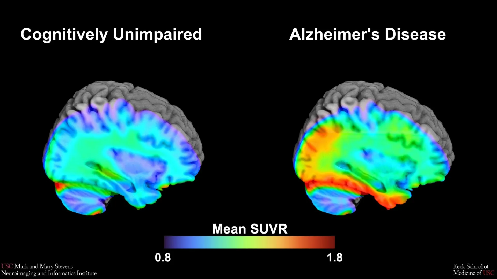
The USC-led study utilized an advanced imaging technology known as tau Positron Emission Tomography (PET). This procedure involves the injection of a specialized radioactive tracer—in this case, a novel agent called 18F-PI-2620—which travels through the bloodstream and binds specifically to tau protein deposits in the brain. When the patient undergoes a PET scan, these tracers "light up," allowing researchers to see exactly where the tau is accumulating and in what volume.
Establishing the "Tau Cut-Point": A New Diagnostic Threshold
The primary objective of the research team, which included over 675 older adults from the HABS-HD cohort, was to establish a "tau cut-point." A cut-point is essentially a mathematical threshold or a "line in the sand." If a patient’s tau levels exceed this specific number, it indicates a high probability of clinically relevant Alzheimer’s pathology. If the levels are below it, the changes may be attributed to normal aging.
Establishing these benchmarks is vital for both clinical trials and future bedside care. Without a standardized cut-point, doctors may struggle to determine which patients are the best candidates for new, tau-targeting therapies. The research team focused their analysis on the medial temporal lobe, a region located deep within the brain that is essential for memory formation and is typically one of the first areas ravaged by tau pathology.
The study’s findings were definitive: when tau levels in the medial temporal lobe exceeded the identified threshold, there was a strong correlation with cognitive impairment. However, the researchers discovered that the efficacy of this benchmark was not universal, revealing a complex interplay between different proteins and different demographic backgrounds.
The Role of Amyloid and the Discovery of Demographic Disparities
One of the most significant revelations of the study was that the tau cut-point was most effective as a predictor of cognitive impairment only when another abnormal protein, amyloid, was also present. This reinforces the "amyloid cascade hypothesis," which suggests that amyloid acts as a catalyst, potentially triggering or accelerating the spread of tau throughout the brain.
Furthermore, the study highlighted a critical gap in current diagnostic models. While the tau cut-point was a highly accurate indicator for Hispanic and non-Hispanic White participants, it did not perform with the same level of reliability for non-Hispanic Black participants.
"Our tau cut-point was able to distinguish whether study participants had cognitive impairment—but only when another abnormal protein, amyloid, was also present… and only in Hispanic and non-Hispanic White participants," noted senior author Meredith N. Braskie, PhD, an assistant professor of neurology at the Keck School of Medicine.
The fact that the benchmark did not perform as expected in non-Hispanic Black participants suggests that the drivers of cognitive decline in this population may involve different biological pathways. This could include vascular issues, such as hypertension or small vessel disease, or other types of proteinopathies that are not yet as well-understood as the amyloid-tau model.
Methodology and the Use of 18F-PI-2620
The technical success of the study is attributed largely to the use of the 18F-PI-2620 tracer. Unlike older tracers, which sometimes suffered from "off-target binding"—where the tracer attaches to things other than tau—18F-PI-2620 is designed for high affinity and high selectivity. This allows for a much cleaner and more accurate measurement of tau in the brain’s most sensitive regions.
The research team analyzed the scans of the 675 participants, categorizing them based on cognitive tests. By comparing the tau levels of those with confirmed cognitive impairment against those who were cognitively healthy, they were able to statistically derive the optimal threshold for the 18F-PI-2620 tracer.
Lead author Victoria R. Tennant, a PhD candidate in USC’s Neuroscience Graduate Program, emphasized that while the study confirms the importance of the medial temporal lobe, it also serves as a call to action. "The limited reliability of tau as an indicator of cognitive impairment in non-Hispanic Black participants highlights the need for more diverse populations in research and for future studies to examine both biological and social determinants of Alzheimer’s disease," Tennant stated.
Chronology of the HABS-HD Study and Collaborative Impact
The Health and Aging Brain Study-Health Disparities (HABS-HD) was launched to address a glaring historical deficit in medical research. For decades, the majority of Alzheimer’s data was gathered from predominantly White, affluent populations. However, statistics show that Hispanic and Black Americans are at a significantly higher risk of developing Alzheimer’s and other dementias.
- 2010s: The realization grows within the scientific community that Alzheimer’s biomarkers may present differently across ethnicities.
- Establishment of HABS-HD: A multi-university collaboration is formed, led by the University of North Texas Health Science Center, to create the most comprehensive database of AD in diverse communities.
- Tracer Development: Next-generation tracers like 18F-PI-2620 are developed and validated, offering better precision than earlier imaging methods.
- Current Study: The Stevens INI team utilizes the HABS-HD cohort to validate these tracers and establish the first diverse-centric tau cut-points.
The study involved a massive collaborative effort, featuring authors from the Alzheimer’s Therapeutic Research Institute at USC, the Waisman Center at the University of Wisconsin-Madison, Washington University School of Medicine in St. Louis, and the University of California, San Francisco.
Broader Implications for Clinical Trials and Personalized Medicine
The implications of this research extend far beyond the laboratory. As the pharmaceutical industry moves toward "precision medicine," having accurate benchmarks for diverse populations is essential for the success of clinical trials. If a drug is designed to clear tau from the brain, researchers must be able to accurately identify who has a "tau-positive" scan. If the current benchmarks only work for certain groups, then other groups risk being excluded from trials or receiving treatments that are not tailored to their specific biological profile.
Arthur W. Toga, PhD, Director of the Stevens INI, highlighted the broader vision of the institute. "This type of imaging is critical for understanding who is at risk and how the disease develops," Toga said. He noted that HABS-HD has already provided vital data on vascular contributions to dementia and the influence of social determinants—such as education, environment, and access to healthcare—on cognitive health.
The study suggests that for some populations, a "one-size-fits-all" biological model of Alzheimer’s may be insufficient. By acknowledging that different pathologies may drive cognitive decline in different groups, the medical community can move toward more personalized care. This might include a broader diagnostic panel that looks at vascular health, inflammation, and multiple protein types, rather than just amyloid and tau.
Analysis of Future Research Directions
The results published in Imaging Neuroscience set the stage for several new avenues of inquiry. First, there is an urgent need to identify the specific "other pathologies" mentioned by Dr. Braskie that may be driving cognitive decline in non-Hispanic Black participants. If tau is not the primary driver, researchers must find out what is.
Second, the study underscores the importance of the medial temporal lobe as a "ground zero" for AD monitoring. Future longitudinal studies—which follow the same individuals over many years—will be necessary to see if these tau cut-points can predict the rate of future decline, not just the current state of impairment.
Finally, the success of the 18F-PI-2620 tracer in this study likely paves the way for its wider adoption in clinical settings. As PET imaging becomes more accessible, these standardized cut-points will provide clinicians with a vital tool for early intervention, potentially allowing patients to begin lifestyle changes or medical treatments years before their symptoms become debilitating.
This research was supported by extensive grants from the National Institute on Aging and the National Institutes of Health, reflecting the federal government’s commitment to solving the Alzheimer’s crisis through the lens of health equity. As the global population ages, the work of the Stevens INI and its collaborators remains at the forefront of ensuring that the benefits of scientific progress reach every community, regardless of ethnic or racial background.