
The Cerebellum: A Focal Point of Multiple Sclerosis Pathology
The cerebellum, located at the back of the brain, is the command center for fine motor skills, posture, and equilibrium. In approximately 80% of multiple sclerosis (MS) cases, this region undergoes significant inflammatory changes. For patients, damage to the cerebellum manifests as tremors, an unsteady gait, and a loss of muscle control known as ataxia. Unlike the temporary relapses seen in early stages of the disease, the symptoms associated with cerebellar damage often become permanent and progressive as healthy tissue is gradually replaced by lesions and scar tissue.
The UC Riverside study, led by Seema Tiwari-Woodruff, a professor of biomedical sciences, and her graduate student Kelley Atkinson, sought to uncover why the cerebellum is so uniquely susceptible to this decline. By examining the interplay between inflammation, demyelination, and cellular metabolism, the researchers identified a critical breakdown in the "powerhouses" of the cell—the mitochondria.
The Mechanics of Neurodegeneration: Beyond Demyelination
For decades, MS research focused primarily on demyelination. In a healthy nervous system, myelin acts like the plastic insulation on an electrical wire, preventing signal leakage and ensuring rapid transmission. When the immune system mistakenly attacks this insulation, signals become sluggish or blocked entirely. However, demyelination alone does not always explain the death of the underlying nerve cells.
The new research suggests that the loss of myelin triggers a secondary, more lethal crisis: mitochondrial dysfunction. Mitochondria are responsible for producing adenosine triphosphate (ATP), the chemical energy that fuels every cellular process. In the cerebellum of MS patients, the researchers observed a significant depletion of a specific mitochondrial protein known as COXIV.
"Our study proposes that inflammation and demyelination in the cerebellum disrupt mitochondrial function, contributing to nerve damage and Purkinje cell loss," Professor Tiwari-Woodruff explained. The absence of COXIV serves as a marker for mitochondrial impairment, suggesting that these cells are essentially starving to death because they can no longer produce the energy required to maintain their complex structures.
The Critical Role of Purkinje Neurons
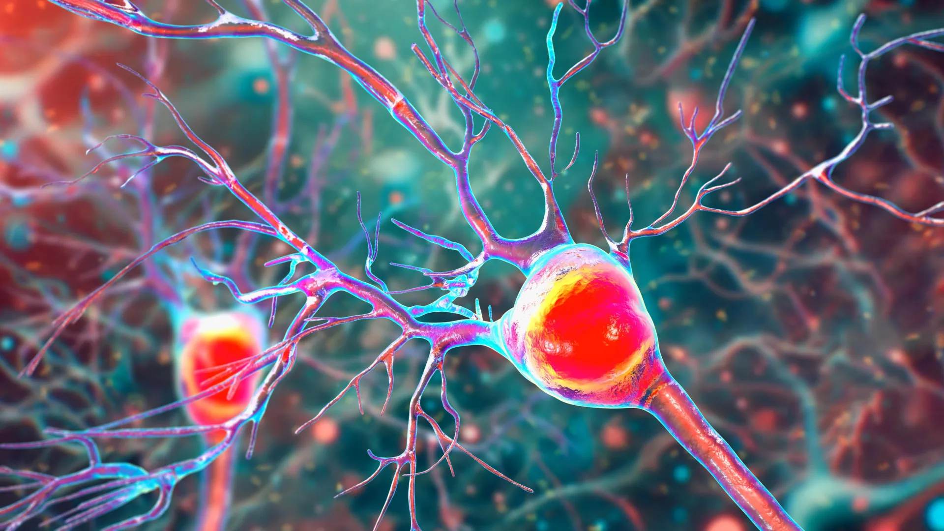
At the heart of the study are Purkinje cells, some of the largest and most metabolically active neurons in the human body. These cells feature intricate, tree-like branches called dendrites that receive thousands of inputs from other parts of the brain. Because of their size and the constant electrical activity they maintain, Purkinje cells have an immense appetite for energy.
When MS-induced inflammation hits the cerebellum, these cells are among the first to suffer. The research team analyzed postmortem cerebellar tissue from individuals who had been diagnosed with secondary progressive MS, comparing it to tissue from healthy donors. The findings were stark: the Purkinje cells in MS-affected brains were not only fewer in number but also showed signs of physical atrophy. They had fewer dendritic branches and lacked the necessary mitochondrial density to sustain their function.
As these neurons disappear, the brain loses its ability to refine motor movements. This leads to the hallmark symptoms of progressive MS, where simple tasks like walking or reaching for an object become increasingly difficult and eventually impossible.
Experimental Insights from the EAE Mouse Model
To validate their findings and observe the chronological progression of the disease, the researchers utilized a mouse model known as experimental autoimmune encephalomyelitis (EAE). This model allows scientists to track the biological changes in the brain in real-time, providing a window into the early stages of the disease that cannot be captured in postmortem human tissue.
The longitudinal study of the EAE mice revealed a specific timeline of decline. In the early stages of the disease, inflammation and the breakdown of myelin were the most prominent features. However, the actual death of the Purkinje cells did not occur immediately. Instead, there was a period of "mitochondrial distress" during which the neurons were still present but were functionally impaired due to failing energy supplies.
"The remaining neurons don’t work as well because their mitochondria start to fail," Tiwari-Woodruff noted. "These problems—less energy, loss of myelin, and damaged neurons—start early, but the actual death of the brain cells tends to happen later, as the disease becomes more severe."
This discovery is significant because it identifies a potential window for therapeutic intervention. If the "energy crisis" can be addressed before the cells actually die, it may be possible to preserve motor function in patients who are in the early or middle stages of MS.
Supporting Data and Statistical Context
The global burden of MS is rising, with the National MS Society reporting that the number of people living with the condition in the United States alone has nearly doubled in recent decades to nearly one million. Approximately 15% of patients are diagnosed with Primary Progressive MS (PPMS) from the onset, while many others transition from Relapsing-Remitting MS (RRMS) to Secondary Progressive MS (SPMS).
The UC Riverside study provides a biological explanation for why progressive forms of the disease are so difficult to treat. While current Disease-Modifying Therapies (DMTs) are highly effective at reducing the frequency of relapses by suppressing the immune system, they have historically been less effective at stopping the slow, "smoldering" neurodegeneration that occurs in the cerebellum. The data from the study suggests that this is because existing treatments do not address the mitochondrial failure occurring within the neurons themselves.
Analysis of Implications: A Shift Toward Neuroprotection
The implications of this research represent a paradigm shift in how MS might be treated in the future. If mitochondrial impairment is a primary driver of cell death, then the next generation of MS drugs may need to focus on "neuroprotection" and "metabolic support" rather than just immunosuppression.
Potential strategies could include:
- Mitochondrial Boosting: Developing compounds that increase the efficiency of the electron transport chain or stabilize proteins like COXIV.
- Early Remyelination: Accelerating the repair of the myelin sheath to reduce the metabolic stress on the underlying axons.
- Metabolic Stabilization: Using antioxidants or metabolic precursors to prevent the oxidative stress that often accompanies mitochondrial failure.
By targeting the health of the mitochondria, clinicians may be able to slow the transition from relapsing-remitting MS to the more severe progressive stages, significantly improving the quality of life for millions of patients.
Future Research: The Role of Support Cells
The UC Riverside team is not stopping at Purkinje cells. Their ongoing research is now expanding to look at other cell types in the cerebellum that may be contributing to the disease environment. This includes oligodendrocytes, the cells responsible for creating myelin, and astrocytes, which provide structural and metabolic support to neurons.
"To answer this, one of our ongoing research projects is focused on studying mitochondria in specific types of brain cells in the cerebellum," Tiwari-Woodruff said. Understanding how these support cells interact with Purkinje neurons during a mitochondrial crisis could reveal even more targets for therapy. For instance, if astrocytes are also experiencing energy failure, they may be unable to provide the nutrients that Purkinje cells need to survive the inflammatory assault.
The Necessity of Sustained Scientific Investment
The study, which was supported by funding from the National Multiple Sclerosis Society and utilized tissue from the NIH NeuroBioBank and the Cleveland Clinic, concludes with a plea for continued support of basic and clinical research.
As the complexity of neurodegenerative diseases becomes more apparent, the need for interdisciplinary research that combines immunology, neurology, and biochemistry is paramount. Professor Tiwari-Woodruff emphasized that medical breakthroughs are the result of long-term investment and that any reduction in scientific funding could stall the development of life-changing treatments.
The findings from the UC Riverside School of Medicine bring the scientific community a step closer to solving one of the most persistent mysteries of MS. By identifying the mitochondrial "engine failure" in the cerebellum, researchers have found a new roadmap for preserving the mobility, independence, and dignity of those living with the disease.