A groundbreaking study led by researchers at the University of California, Riverside (UCR) and published in the Proceedings of the National Academy of Sciences (PNAS) has shed light on the cellular mechanisms responsible for this decline. The research points to the malfunctioning of mitochondria—the specialized organelles responsible for energy production—as a major driver in the breakdown of Purkinje cells. These cells are the primary output neurons of the cerebellar cortex, and their survival is paramount to preserving motor function in MS patients.
The Pathological Intersection of Inflammation and Demyelination
To understand the significance of the UCR findings, one must first look at the traditional understanding of MS pathology. The disease is defined by two primary processes: chronic inflammation and demyelination. Demyelination occurs when the body’s immune system mistakenly attacks the myelin sheath, the fatty, insulating layer that wraps around nerve fibers (axons). Much like the plastic coating on an electrical wire, myelin ensures that electrical impulses travel rapidly and efficiently between the brain and the rest of the body.
When this insulation is stripped away, the transmission of nerve signals becomes sluggish or is blocked entirely. However, the UCR study suggests that demyelination does more than just slow down signals; it creates a hostile environment that disrupts the internal machinery of the nerve cells themselves.
Mitochondria, often described as the "powerhouses" of the cell, are responsible for generating adenosine triphosphate (ATP), the chemical energy required for almost every cellular function. In the context of MS, the research team discovered that the double-hit of inflammation and myelin loss leads to a catastrophic failure of these energy producers.
"Our study proposes that inflammation and demyelination in the cerebellum disrupt mitochondrial function, contributing to nerve damage and Purkinje cell loss," explained Seema Tiwari-Woodruff, a professor of biomedical sciences in the UC Riverside School of Medicine and the study’s lead investigator. The team observed a significant reduction in a specific mitochondrial protein known as COXIV within demyelinated Purkinje cells. This deficiency suggests that the cells are essentially starving for energy, leading to a state of metabolic exhaustion that eventually triggers cell death.
The Vital Role of Purkinje Neurons in Motor Coordination
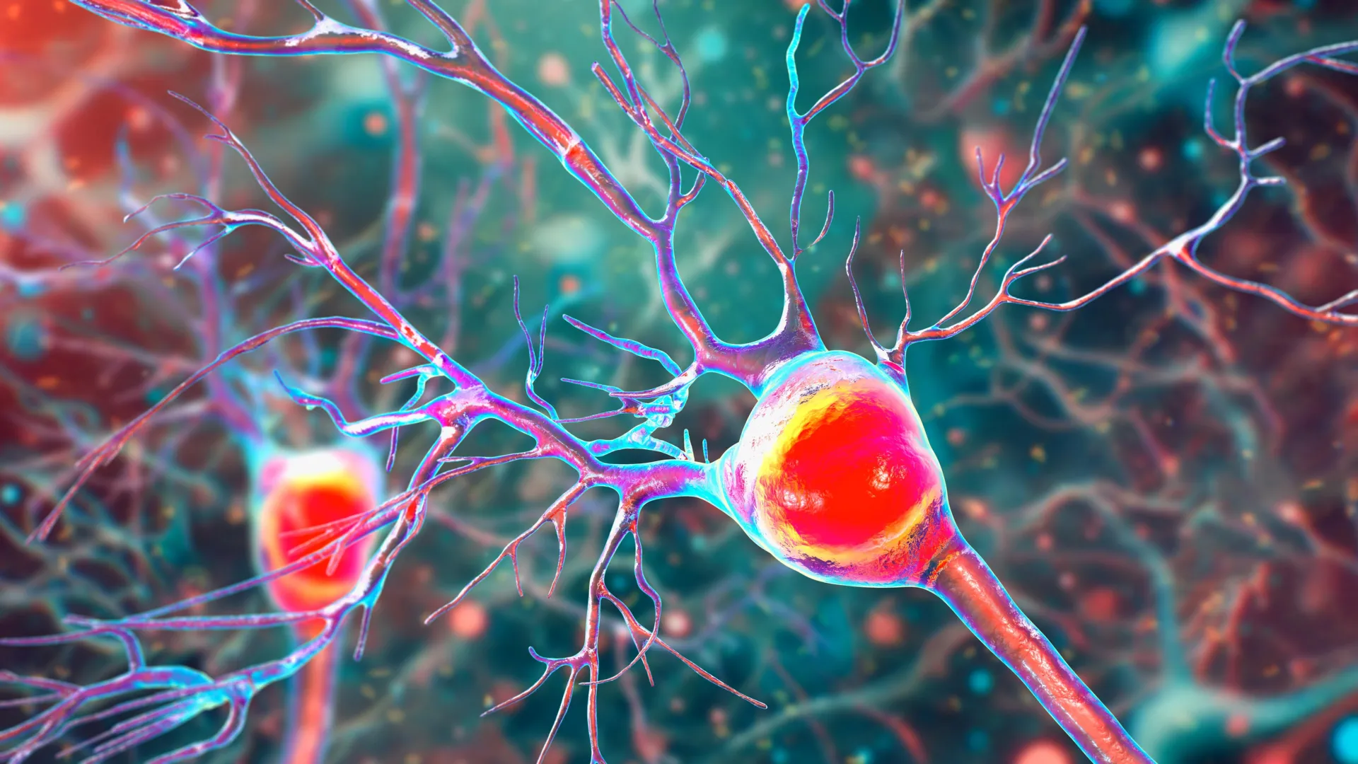
The cerebellum serves as the body’s internal "flight controller," processing sensory input to fine-tune motor activity. At the heart of this system are Purkinje neurons. These are among the largest and most complex cells in the human brain, characterized by an extensive, tree-like array of dendrites that allow them to receive and integrate massive amounts of information.
"Inside the cerebellum are special cells called Purkinje neurons," Tiwari-Woodruff said. "These large, highly active cells help coordinate smooth, precise movements—like dancing, throwing a ball, or even just walking. They’re essential for balance and fine motor skills."
Because Purkinje cells are so metabolically active, they have high energy demands. This makes them particularly vulnerable to mitochondrial impairment. In MS and related neurodegenerative disorders, the gradual death of these cells leads to a condition known as ataxia. Ataxia is characterized by a lack of voluntary coordination of muscle movements, which can manifest as difficulty swallowing, impaired speech, and an unstable, wide-based walking pattern.
The research team analyzed postmortem cerebellar tissue from individuals who had been diagnosed with secondary progressive MS, a stage of the disease where disability increases steadily. By comparing this tissue with samples from healthy donors—sourced from the National Institutes of Health’s NeuroBioBank and the Cleveland Clinic—the researchers were able to visualize the physical toll of the disease. They found that in MS patients, Purkinje neurons exhibited fewer branches (dendritic shrinkage), significant myelin loss, and a clear depletion of mitochondrial markers.
Insights from the EAE Mouse Model
To further validate their findings and observe the progression of the disease in real-time, the researchers utilized an experimental autoimmune encephalomyelitis (EAE) mouse model. EAE is a widely accepted laboratory proxy for MS, as it mimics the inflammatory and demyelinating characteristics of the human condition.
By tracking the mice over the course of the disease, the team was able to establish a clear timeline of neurodegeneration. They observed that myelin breakdown occurs early in the disease process, followed closely by the onset of mitochondrial dysfunction. Interestingly, while the "energy crisis" within the cells begins early, the actual death of the neurons tends to occur in the later, more severe stages of the disease.
"The remaining neurons don’t work as well because their mitochondria, the energy-producing parts, start to fail," Tiwari-Woodruff noted. "These problems—less energy, loss of myelin, and damaged neurons—start early, but the actual death of the brain cells tends to happen later. The loss of energy in brain cells seems to be a key part of what causes damage in MS."
This chronological insight is vital for the development of new therapies. It suggests a "window of opportunity" where medical intervention could potentially stabilize mitochondrial health and prevent the irreversible loss of Purkinje cells, even after demyelination has begun.
Toward New Mitochondrial-Targeted Therapies
Current MS treatments largely focus on modulating the immune system to reduce the frequency of inflammatory "flares" or relapses. While these disease-modifying therapies (DMTs) are effective at slowing the progression of relapsing-remitting MS, they have shown limited success in stopping the neurodegeneration associated with progressive forms of the disease.
The UCR study shifts the focus toward neuroprotection—specifically, protecting the metabolic integrity of the neurons. "Our findings offer critical insights into the progression of cerebellar dysfunction in MS," Tiwari-Woodruff said. "Targeting mitochondrial health may represent a promising strategy to slow or prevent neurological decline and improve quality of life for people living with MS."
Potential future treatments could involve compounds that boost mitochondrial biogenesis (the creation of new mitochondria), antioxidants that protect mitochondria from oxidative stress, or metabolic precursors that help cells maintain ATP production despite the loss of myelin. By addressing the "energy failure" identified in the study, clinicians may eventually be able to preserve mobility and coordination in patients who currently have few options.
Expanding the Scope: Glial Cells and Future Research
The UCR research team, which included graduate student Kelley Atkinson and several other contributors—Shane Desfor, Micah Feria, Maria T. Sekyia, Marvellous Osunde, Sandhya Sriram, Saima Noori, Wendy Rincón, and Britany Bello—is not stopping at Purkinje cells. Their ongoing work aims to determine if mitochondrial failure is a localized issue or a widespread phenomenon affecting other cell types in the cerebellum.
One area of particular interest is the role of glial cells, such as oligodendrocytes and astrocytes. Oligodendrocytes are the cells responsible for producing myelin, while astrocytes provide structural and metabolic support to neurons. If mitochondrial damage is also occurring in these support cells, it could create a "vicious cycle" where the brain’s ability to repair itself is also compromised.
"To answer this, one of our ongoing research projects is focused on studying mitochondria in specific types of brain cells in the cerebellum," Tiwari-Woodruff stated. "Such research can open the door to finding ways to protect the brain early on—like boosting energy in brain cells, helping them repair their protective myelin coating, or calming the immune system before too much damage is done."
The Global Implications of Sustained Scientific Investment
The study, funded by the National Multiple Sclerosis Society, underscores the necessity of long-term investment in basic and translational science. As the global population ages and the prevalence of autoimmune disorders remains high, understanding the fundamental biology of neurodegeneration is essential for public health.
Tiwari-Woodruff emphasized that scientific progress is a cumulative effort that requires consistent support. "Cutting funding to science only slows progress when we need it most," she said. "Public support for research matters now more than ever."
As the medical community moves toward a more nuanced understanding of MS—one that looks beyond inflammation to the metabolic health of individual neurons—the findings from UC Riverside provide a vital roadmap. For the millions of people living with MS, particularly those struggling with the loss of balance and movement, these insights offer a new sense of hope for treatments that do more than just manage symptoms, but actually protect the brain’s ability to function.