The study is a cornerstone of the Health and Aging Brain Study-Health Disparities (HABS-HD), a comprehensive, multi-university collaboration led by the University of North Texas Health Science Center and supported by the National Institute on Aging (NIA). By analyzing a cohort of over 675 older adults, the USC-led team sought to pinpoint the exact threshold of tau protein—a hallmark of Alzheimer’s—that distinguishes normal aging from clinically relevant cognitive impairment.
The Biological Mechanics of Alzheimer’s Disease
To understand the significance of this benchmark, it is essential to contextualize the roles of two primary proteins in the progression of Alzheimer’s disease: amyloid-beta and tau. For decades, the "amyloid hypothesis" dominated research, suggesting that the buildup of amyloid plaques between neurons was the primary driver of the disease. However, recent advancements have shifted the focus toward tau protein, which forms "tangles" inside the neurons themselves.
While amyloid plaques often appear years before clinical symptoms manifest, the accumulation of tau tangles is more closely correlated with the actual onset of memory loss and cognitive decline. Tau disrupts the internal transport system of neurons, leading to cell death and brain atrophy. The ability to accurately measure and categorize tau levels in a living brain is therefore paramount for early diagnosis and the development of targeted therapies.
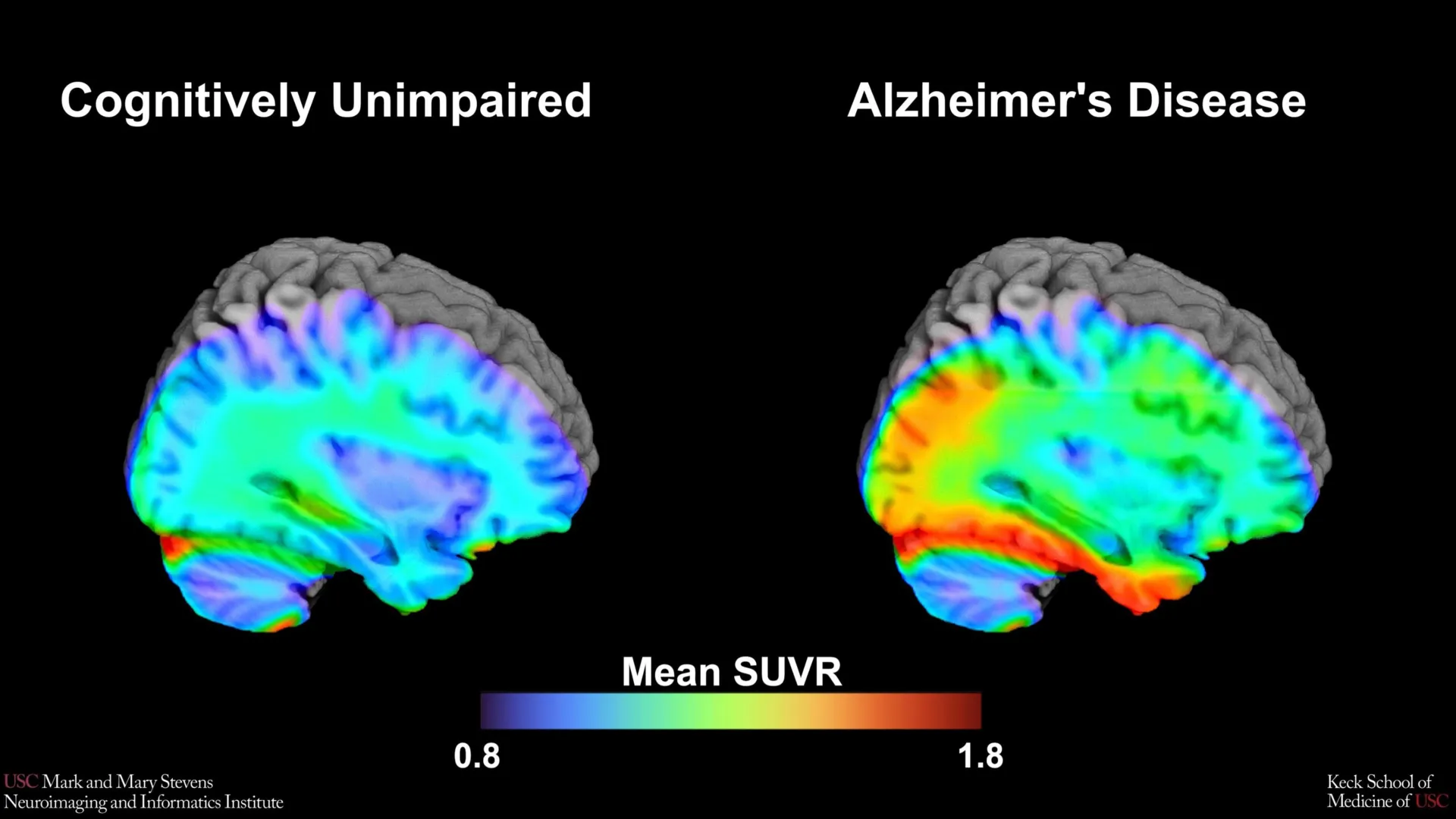
The USC study utilized a cutting-edge imaging tracer known as 18F-PI-2620. This specialized radioactive agent binds to tau proteins in the brain, allowing them to be visualized and quantified through a tau PET scan. By measuring the "signal" from this tracer, researchers can determine the density and location of tau accumulation.
Methodology and the Identification of Tau Cut-Points
The research team analyzed data from 675 participants enrolled in the HABS-HD study, a group purposefully selected to represent the diversity of the aging American population. The participants underwent rigorous cognitive testing and neuroimaging to correlate their mental health status with biological markers.
The primary objective was to establish "tau cut-points"—specific numerical thresholds of protein density that indicate a high risk for Alzheimer’s or early-stage cognitive impairment. The researchers focused their analysis on the medial temporal lobe, a region deep within the brain that is essential for memory formation and is typically one of the first areas affected by tau pathology.
The findings revealed that when tau levels in the medial temporal lobe exceeded a specific threshold, it served as a strong indicator of cognitive impairment. However, this benchmark was not universal. The researchers discovered that the tau cut-point was highly effective at identifying impairment only when another condition was met: the presence of amyloid plaques. This "dual-protein" requirement underscores the synergistic nature of Alzheimer’s pathology, where the interaction between amyloid and tau appears to accelerate neurodegeneration.
Disparities in Diagnostic Accuracy Across Ethnic Groups
One of the most significant findings of the study—and one that carries profound implications for the future of health equity—is the variation in how these benchmarks performed across different racial and ethnic groups. The established tau cut-point was found to be a reliable indicator of cognitive impairment for Hispanic and non-Hispanic White participants. However, it did not perform as expected for non-Hispanic Black participants.
In the non-Hispanic Black cohort, the correlation between this specific tau threshold and cognitive decline was less clear. This suggests that the biological drivers of Alzheimer’s and related dementias may differ across populations. Lead author Victoria R. Tennant, a PhD candidate in USC’s Neuroscience Graduate Program, noted that the limited reliability of tau as a singular indicator in Black participants highlights a critical gap in current medical understanding.
"This suggests that other pathologies or conditions may be driving cognitive decline in this group," explained senior author Meredith N. Braskie, PhD, assistant professor of neurology. These other factors could include vascular health issues, such as hypertension or small vessel disease, or different inflammatory responses that are not captured by a standard tau PET scan.
The HABS-HD Framework and the Chronology of the Study
The Health and Aging Brain Study-Health Disparities (HABS-HD) was launched to address the historical lack of diversity in Alzheimer’s research. Historically, the vast majority of clinical trials and neuroimaging studies have been conducted on non-Hispanic White populations, leading to diagnostic tools and treatments that may not be equally effective for all.
Since its inception, HABS-HD has evolved into the most comprehensive study of its kind. The collaboration involves several top-tier institutions, including the University of North Texas Health Science Center, USC, the University of Wisconsin-Madison, Washington University in St. Louis, and the University of California, San Francisco.
The chronology of this specific study began with the recruitment of a diverse cohort, followed by the administration of the 18F-PI-2620 tracer in clinical settings. Over several years, the data from these scans were cross-referenced with longitudinal cognitive assessments. The publication of these findings in Imaging Neuroscience marks a pivotal moment in the project’s timeline, shifting the focus from data collection to the practical application of diagnostic benchmarks.
Supporting Data and Technical Analysis
The use of the 18F-PI-2620 tracer represents a technical leap forward. Older tracers often suffered from "off-target binding," where the tracer would stick to proteins other than tau, creating "noise" in the data. The 18F-PI-2620 tracer is designed for higher specificity, allowing for a cleaner signal in the medial temporal lobe.
Data from the study showed that the medial temporal lobe’s tau levels were the most predictive of cognitive status, confirming its role as a "ground zero" for Alzheimer’s progression. However, the requirement for amyloid positivity suggests that tau PET scans should perhaps not be interpreted in isolation. For clinicians, this means that a "positive" tau scan might only be clinically actionable if an amyloid PET scan or a cerebrospinal fluid (CSF) test also confirms the presence of amyloid plaques.
For Hispanic participants, the study’s findings are particularly vital. Statistics from the Alzheimer’s Association indicate that Hispanics are about 1.5 times as likely as older Whites to have Alzheimer’s or other dementias. By validating a specific tau cut-point for this group, the USC researchers have provided a tool that could lead to earlier intervention and better management of the disease in a high-risk population.
Official Responses and Clinical Implications
The broader scientific community has reacted to these findings with a mix of optimism and a call for further investigation into health disparities. Arthur W. Toga, PhD, director of the Stevens INI, emphasized the importance of the study’s focus on diverse communities.
"This type of imaging is critical for understanding who is at risk and how the disease develops," Toga said. "HABS-HD has already produced key findings related to ethnic variations in AD biomarkers, the influences of social determinants on cognitive health, and vascular contributions to dementia. We hope this work will lead to more personalized care and better outcomes for all communities."
The clinical implications are twofold. First, the study provides a standardized "language" for interpreting 18F-PI-2620 PET scans, which can help clinicians move away from subjective interpretations of "high" or "low" tau. Second, it serves as a cautionary tale against a "one-size-fits-all" approach to neurology. If a diagnostic marker works for one group but not another, the medical community must investigate why—whether the cause is genetic, environmental, or related to social determinants of health.
Future Directions: Beyond Biological Markers
The USC study opens several new avenues for research. One priority is investigating why the tau cut-point was less effective for non-Hispanic Black participants. Future studies within the HABS-HD framework will likely examine the intersection of tau pathology with vascular risk factors, which are known to be higher in Black communities due to systemic health disparities.
Furthermore, the research highlights the need for "multi-modal" diagnostics. In the future, a patient’s risk profile might be determined by a combination of tau PET scans, amyloid tests, genetic screening (such as the APOE-ε4 allele), and an assessment of social factors like education level, diet, and exposure to environmental pollutants.
The goal is the realization of "precision medicine" in neurology—the ability to tailor a treatment plan to an individual’s specific biological and demographic profile. As new Alzheimer’s drugs that target amyloid and tau (such as lecanemab and donanemab) become more widely available, having accurate benchmarks to identify the right patients for these treatments is more critical than ever.
The work of the Stevens INI and the HABS-HD collaborators ensures that as the medical community moves toward a cure for Alzheimer’s, no population is left behind. By refining how we see the disease through the lens of advanced imaging, researchers are paving the way for a future where Alzheimer’s can be caught early, understood deeply, and treated effectively for everyone.