The Mechanics of Neuro-Waste Clearance
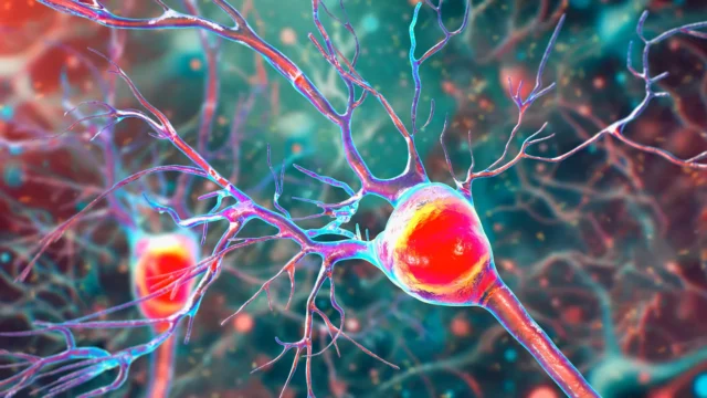
The human brain is an incredibly active organ, consuming a disproportionate amount of the body’s energy and, consequently, producing a significant volume of metabolic waste. To maintain health, the brain utilizes a specialized drainage architecture known as the glymphatic system. At the heart of this system are perivascular spaces—minute, fluid-filled channels surrounding the brain’s blood vessels. These channels act as the primary conduits for flushing out toxic proteins, most notably beta-amyloid and tau.
In a healthy brain, these proteins are efficiently cleared into the cerebrospinal fluid and eventually exit the central nervous system. However, when this drainage system becomes inefficient or blocked, the perivascular spaces begin to dilate and enlarge. This study confirms that these enlarged spaces are not merely incidental findings but are directly correlated with the accumulation of the "plaques and tangles" that define Alzheimer’s pathology. By the time these spaces become visible on an MRI, it indicates a "clogged" state where the brain is no longer able to keep up with the disposal of harmful substances, leading to cellular damage and cognitive impairment.
Addressing the Research Gap in Asian Demographics
One of the most significant aspects of the NTU Singapore study is its focus on Asian populations, a demographic that has historically been underrepresented in global Alzheimer’s research. For decades, the majority of clinical data regarding dementia has been derived from Caucasian cohorts in North America and Europe. This geographic bias has created gaps in medical understanding, as genetic and environmental factors vary significantly across different ethnic groups.
Associate Professor Nagaendran Kandiah, Director of the Dementia Research Centre (Singapore) at LKCMedicine and lead investigator of the study, highlighted the necessity of region-specific data. He pointed out that the genetic landscape of Alzheimer’s in Asia differs markedly from that in the West. For instance, the apolipoprotein E4 (APOE4) gene is the most well-known genetic risk factor for Alzheimer’s. While approximately 50 to 60 percent of Caucasians with dementia carry this gene, it is found in less than 20 percent of Singaporean dementia patients.
By examining nearly 1,000 participants in Singapore—comprising a representative mix of Chinese, Malay, and Indian ethnicities—the NTU team has provided a more accurate blueprint for how the disease progresses in Asian brains. This inclusive approach ensures that diagnostic tools developed from the research are culturally and genetically relevant to billions of people across the continent.
Methodology: Comparing Brain Scans and Blood Biomarkers
The study’s methodology was rigorous, involving a multi-modal approach that combined advanced neuroimaging with blood-based biochemical analysis. The research team categorized the participants into two primary groups: approximately 350 individuals with normal cognitive function and the remainder showing signs of mild cognitive impairment (MCI). MCI is often considered a "prodromal" or precursor stage to full-blown dementia, where individuals experience noticeable memory lapses but can still perform daily activities.
The researchers utilized MRI scans to identify and quantify the presence of enlarged perivascular spaces. Simultaneously, they measured seven specific Alzheimer’s-related biochemicals in the participants’ blood. These markers included various forms of beta-amyloid and tau proteins, which serve as "liquid biopsies" for the state of the brain.
The results were striking. Enlarged perivascular spaces were significantly more prevalent in the MCI group than in the healthy control group. Furthermore, these enlarged spaces were strongly linked to four out of the seven blood-based biochemical markers. This correlation provides robust evidence that the "clogged drains" observed on the MRI are a direct reflection of the underlying molecular pathology of Alzheimer’s, including the buildup of amyloid plaques and tau tangles.
EPVS vs. White Matter Damage: A Superior Early Marker
Traditionally, clinicians have looked at white matter hyperintensities—areas of damage to the brain’s "wiring" caused by reduced blood flow—as a primary indicator of vascular-related cognitive decline. While white matter damage is easily identifiable on MRIs and remains a useful diagnostic tool, the NTU study found that enlarged perivascular spaces (EPVS) might actually be a more sensitive indicator for the very earliest stages of Alzheimer’s.
Among participants with mild cognitive impairment, the statistical link between Alzheimer’s biochemicals and EPVS was notably stronger than the link between those same biochemicals and white matter damage. This discovery suggests that the failure of the brain’s waste removal system may precede the more overt structural damage seen in white matter. Consequently, EPVS could offer a narrower window for early intervention, allowing doctors to identify at-risk patients years before traditional markers become severe.
Clinical Reactions and the Synergy of Brain Health
The findings have drawn praise from the wider medical community in Singapore. Dr. Rachel Cheong Chin Yee, a Senior Consultant at Khoo Teck Puat Hospital’s Department of Geriatric Medicine, noted that the study underscores the intricate relationship between the brain’s vascular health and neurodegeneration. She emphasized that identifying these markers before symptoms appear is the "holy grail" of dementia care, as it allows for preventative strategies that could preserve quality of life for longer periods.
Similarly, Dr. Chong Yao Feng, a Consultant at the National University Hospital’s Division of Neurology, pointed out that the study challenges the traditional separation of cerebrovascular disease and Alzheimer’s disease. For years, these were treated as distinct paths to dementia. However, the NTU research demonstrates a synergistic interaction: when the "plumbing" (the vascular system) fails, it accelerates the "pollution" (Alzheimer’s proteins), which in turn further damages the brain. Dr. Chong advised that clinicians reviewing routine MRIs should now view EPVS with a higher degree of suspicion, recognizing them as potential precursors to Alzheimer’s rather than just benign signs of aging.
The Path to Early Intervention and Treatment
The practical implications of this research are profound. Current gold-standard tests for Alzheimer’s, such as Positron Emission Tomography (PET) scans or lumbar punctures to analyze cerebrospinal fluid, are often expensive, invasive, or unavailable in primary care settings. In contrast, MRI scans are already a routine part of the clinical workup for patients reporting memory issues.
"Since these brain anomalies can be visually identified on routine MRI scans performed to evaluate cognitive decline, identifying them could complement existing methods to detect Alzheimer’s earlier, without having to do and pay for additional tests," said Assoc Prof Kandiah.
Early detection is the cornerstone of modern Alzheimer’s management. While a definitive cure remains elusive, early identification allows for the implementation of lifestyle interventions—such as blood pressure management, cognitive training, and diet modifications—that are most effective when started in the prodromal phase. Furthermore, as new disease-modifying therapies emerge, identifying the right candidates early will be essential for treatment success.
Future Outlook: A Longitudinal Vision
The NTU research team, including first author and medical student Justin Ong, is not stopping at this cross-sectional analysis. The next phase of the research involves a longitudinal study, where the participants will be tracked over several years. This will allow the scientists to observe the direct trajectory from the appearance of enlarged perivascular spaces to the clinical diagnosis of Alzheimer’s dementia.
If the longitudinal data confirms that EPVS is a reliable predictor of future decline, it could lead to the development of standardized scoring systems for MRI scans. This would provide radiologists and neurologists with a clear metric to assess a patient’s risk profile during a standard check-up.
As the global population ages, particularly in Asia where the number of dementia cases is expected to soar in the coming decades, the search for accessible diagnostic markers is more urgent than ever. The discovery of the link between the brain’s "clogged drains" and Alzheimer’s pathology represents a significant step forward in the quest to map the earliest footprints of a devastating disease, offering hope for earlier intervention and better outcomes for millions.