
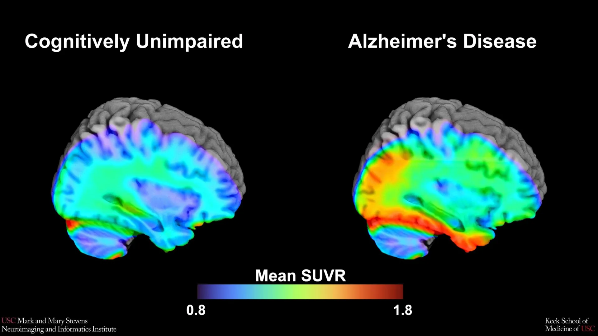
This research is a cornerstone of the Health and Aging Brain Study-Health Disparities (HABS-HD), a massive multi-university collaborative effort. Led by the University of North Texas Health Science Center (UNTHSC) and supported by the National Institute on Aging (NIA), the HABS-HD initiative represents one of the most comprehensive attempts to date to understand the ethnic and racial variations in dementia. By analyzing the neurological profiles of a diverse cohort, the USC-led team is challenging the "one-size-fits-all" approach to Alzheimer’s diagnostics that has dominated the field for decades.
The Science of Tau PET Imaging and the 18F-PI-2620 Tracer
To understand the significance of this benchmark, it is necessary to examine the biological hallmarks of Alzheimer’s disease. For years, the primary focus of AD research was amyloid-beta plaques—protein fragments that accumulate between neurons. However, modern neurology has increasingly shifted its focus toward tau, a protein that forms "tangles" inside the neurons themselves. While amyloid may appear years before symptoms manifest, the accumulation of tau is more closely correlated with actual cognitive decline and the death of brain cells.
The research team utilized an advanced imaging technique known as Positron Emission Tomography (PET) using a specific radioactive tracer called 18F-PI-2620. This tracer is designed to bind specifically to aggregated tau proteins in the brain. When a patient undergoes a tau PET scan, the tracer highlights areas of high tau density, allowing researchers to visualize the "topography" of the disease.
The primary goal of the USC study was to establish "tau cut-points." In medical terms, a cut-point is a threshold value used to categorize a result as either positive or negative. By identifying the exact level of tau accumulation that signals the transition from healthy aging to early-stage Alzheimer’s, the team has provided a metric that can be used to standardize how scans are interpreted across different clinical and research settings.
Methodology: Analyzing a Diverse Cohort
The study involved a rigorous analysis of over 675 older adults enrolled in the HABS-HD study. This cohort is unique due to its intentional inclusion of diverse populations, which are historically underrepresented in clinical trials. The participants underwent comprehensive cognitive testing to determine their level of mental impairment, as well as PET scans to measure both amyloid and tau levels.
The researchers focused their analysis on the medial temporal lobe, a region located deep within the brain that is essential for memory formation and is typically one of the first areas affected by tau pathology. By comparing the PET scans of cognitively impaired individuals with those of cognitively healthy participants, the team sought to find a mathematical threshold where tau levels consistently predicted impairment.
The findings confirmed that when tau levels in the medial temporal lobe exceeded a specific threshold, it was a strong indicator of Alzheimer’s-related cognitive impairment. However, the data also revealed a complex relationship between different proteins. The tau cut-point was most accurate in predicting cognitive decline only when amyloid plaques were also present in the brain. This "dual-protein" requirement underscores the prevailing theory that amyloid may act as a catalyst that accelerates the spread of tau tangles throughout the cortex.
Racial Disparities and the "Black Participant Paradox"
One of the most significant and sobering findings of the study was the failure of the tau cut-point to accurately predict cognitive impairment in non-Hispanic Black participants. While the benchmark was a reliable indicator for Hispanic and non-Hispanic White groups, the same levels of tau did not correlate with the same levels of cognitive decline in Black participants.
"In non-Hispanic Black participants, the tau cut-point did not perform as expected," noted senior author Meredith N. Braskie, PhD, an assistant professor of neurology at the Keck School of Medicine. "This suggests that other pathologies or conditions may be driving cognitive decline in this group."
This discovery has profound implications for the future of Alzheimer’s care. It suggests that the biological "signature" of dementia may vary across different racial and ethnic groups. For instance, Black populations in the United States have higher rates of vascular risk factors, such as hypertension and diabetes, which can lead to vascular dementia—a condition driven by poor blood flow to the brain rather than protein accumulation. If clinicians rely solely on tau PET scans calibrated for White populations, they risk misdiagnosing or overlooking the primary drivers of cognitive loss in Black patients.
A Chronology of Progress in Alzheimer’s Biomarkers
The establishment of this new benchmark is part of a broader timeline of evolution in Alzheimer’s diagnostics.
- Pre-2000s: Alzheimer’s could only be definitively diagnosed through an autopsy after a patient’s death. Clinical diagnosis was based on the exclusion of other diseases.
- Early 2000s: The development of the first amyloid PET tracers allowed researchers to see plaques in living patients for the first time.
- 2010s: Tau PET tracers were developed, providing a better map of symptomatic progression.
- 2020s: The focus shifted toward "biomarker-based" diagnosis, where the presence of specific proteins (amyloid and tau) defines the disease, even in the absence of symptoms.
- Present: Studies like the USC tau benchmark are moving the field toward "precision medicine," where diagnostic thresholds are adjusted for the individual’s demographic and genetic background.
Perspectives from the Research Leadership
The leadership at the Stevens INI emphasizes that these findings are not just academic but are essential for the development of effective treatments. Arthur W. Toga, PhD, director of the Stevens INI, highlighted the broader mission of the HABS-HD study.
"This type of imaging is critical for understanding who is at risk and how the disease develops," Toga stated. "HABS-HD has already produced key findings related to ethnic variations in AD biomarkers, the influences of social determinants on cognitive health, and vascular contributions to dementia. We hope this work will lead to more personalized care and better outcomes for all communities."
Lead author Victoria R. Tennant, a PhD candidate in USC’s Neuroscience Graduate Program, emphasized the need for a more holistic view of the disease. While the medial temporal lobe tau is a powerful indicator, the study’s failure to find the same reliability in Black participants points toward the necessity of examining both biological and social determinants of health. These social determinants include long-term exposure to environmental toxins, chronic stress related to systemic inequality, and disparities in access to preventative healthcare—all of which can influence brain health in ways that a protein scan cannot fully capture.
Implications for Clinical Trials and Future Treatment
The identification of these cut-points is expected to have an immediate impact on the design of clinical trials. Currently, many pharmaceutical companies developing anti-tau drugs use tau PET scans to screen participants. If the thresholds used to "enroll" patients are based on data that does not apply to Black populations, these trials will continue to lack diversity, and the resulting drugs may not be effective for all groups.
Furthermore, the study highlights the importance of multi-modal diagnostics. Relying on a single biomarker (like tau) may provide an incomplete picture. Future diagnostic protocols will likely need to combine tau PET scans with amyloid scans, vascular health assessments, and perhaps even blood-based biomarkers to provide a comprehensive risk profile.
Broadening the Scope of Alzheimer’s Research
The USC study is part of a growing movement to democratize neuroscience. Historically, up to 90% of participants in Alzheimer’s clinical trials have been of European descent. This has created a "knowledge gap" that leaves minority populations—who are often at a higher risk for dementia—without validated diagnostic tools.
By establishing that tau cut-points work differently across populations, the USC team has provided a roadmap for future research. The next steps for the HABS-HD collaboration will involve investigating the specific "other pathologies" mentioned by Dr. Braskie. This includes looking at TDP-43 (another protein associated with dementia), microvascular lesions, and the role of inflammation in the brain.
The research was supported by a diverse array of institutions, including the Alzheimer’s Therapeutic Research Institute at USC, the Waisman Center at the University of Wisconsin-Madison, and Washington University School of Medicine in St. Louis. This level of institutional support underscores the scientific community’s recognition that the future of Alzheimer’s research must be inclusive to be successful.
As the global population ages, the burden of Alzheimer’s disease is expected to triple by 2050. The work being done at the Stevens INI ensures that as new treatments and diagnostic tools emerge, they are calibrated for the reality of a diverse global population, ensuring that no community is left behind in the fight against neurodegenerative disease.