
The human vestibular system is a complex network of sensory organs and neural pathways responsible for maintaining balance, spatial orientation, and gaze stability. When the vestibular nerve—the primary conduit for this vital information—is compromised, the result is often a profound and debilitating loss of equilibrium. While many patients experience a significant recovery following an acute vestibular event, a phenomenon known as "central compensation" often leaves a lingering sensitivity. This residual vulnerability means that even years after the initial injury, seemingly routine activities, such as receiving a massage or maintaining specific head positions, can trigger a recurrence of symptoms. Understanding the mechanisms of the vestibular nerve, the role of the musculoskeletal system, and the influence of psychological stress is essential for managing the long-term recovery of those affected by these invisible yet life-altering conditions.
The Anatomy and Physiology of the Vestibular Nerve
The vestibular nerve, also known as the eighth cranial nerve (CN VIII), serves as the communication bridge between the inner ear and the brain. It is divided into two primary branches: the vestibular portion, which handles balance, and the cochlear portion, which facilitates hearing. Within the inner ear, specialized structures known as the semicircular canals and otolith organs detect rotational movement and linear acceleration, respectively. These organs convert mechanical energy from head movement into electrical signals, which the vestibular nerve then transmits to the vestibular nuclei in the brainstem and the cerebellum.
The brain processes these signals to coordinate the Vestibulo-Ocular Reflex (VOR), which keeps vision stable during head movement, and the Vestibulospinal Reflex (VSR), which maintains upright posture. When the nerve is "tweaked" or damaged—clinically referred to as vestibular neuritis or labyrinthitis—the flow of information becomes asymmetrical. The brain receives conflicting data: one ear signals movement while the other remains silent. This sensory mismatch is the root cause of vertigo, the spinning sensation that often leaves patients incapacitated during the acute phase of the disorder.
Chronology of Vestibular Injury and Recovery
The trajectory of a vestibular injury typically follows three distinct phases: the acute phase, the compensation phase, and the maintenance phase.
During the acute phase, which can last from several days to a few weeks, the patient often experiences severe vertigo, nausea, and an inability to walk or stand without assistance. This is usually caused by inflammation of the nerve, often attributed to viral infections or, less commonly, vascular ischemia (reduced blood flow). In this stage, medical intervention focuses on suppressing the vestibular system with medications to reduce the intensity of the spinning.
The second phase, central compensation, is where the brain begins to adapt. Because the vestibular nerve may not fully regain its original function, the brain must "re-calibrate." It learns to rely more heavily on visual cues and proprioceptive input (sensory information from the muscles and joints) to make up for the deficit in the inner ear. This phase can take weeks or months and is often facilitated by Vestibular Rehabilitation Therapy (VRT).
The final stage is the maintenance or "mostly better" phase. At this point, the patient can function normally in daily life but may experience "decompensation." This occurs when the brain’s compensatory mechanisms are overwhelmed by fatigue, illness, or unusual sensory environments. This explains why a patient who has recovered may suddenly feel dizzy again when placed in a face-down position for an hour during a massage; the brain, deprived of visual cues and placed in a static, unusual posture, struggles to maintain the delicate balance it has worked so hard to achieve.
Supporting Data: Prevalence and Economic Impact
According to data from the National Institute on Deafness and Other Communication Disorders (NIDCD), approximately 35% of adults in the United States aged 40 years and older—roughly 69 million people—have experienced some form of vestibular dysfunction. The prevalence increases significantly with age. Furthermore, vestibular disorders are a leading cause of falls in the elderly, which account for a substantial portion of emergency room visits and healthcare expenditures.
Research published in the Journal of Vestibular Research indicates that chronic vestibular symptoms contribute to significant economic loss. Patients often require extended leaves from work, and many report a permanent decrease in productivity due to "brain fog" and motion sensitivity. The psychological toll is equally measurable; studies show that up to 50% of patients with chronic vestibular issues develop secondary anxiety or panic disorders, as the constant threat of a vertigo spell creates a state of perpetual hyper-vigilance.

The Role of Muscle Tightness and the Cervicogenic Connection
A frequent concern among patients is whether physical tension or spinal alignment can damage the vestibular nerve. Clinically, it is rare for a vertebra to directly compress the vestibular nerve, as the nerve is protected within the bony structure of the skull. However, the relationship between the neck (the cervical spine) and the balance system is profound.
This relationship is known as the cervicogenic component of balance. The muscles of the neck are densely packed with proprioceptors that tell the brain exactly where the head is in relation to the body. When a patient experiences a vestibular injury, they often stiffen their neck muscles instinctively to "lock" their head in place and minimize dizziness. This chronic guarding leads to muscle shortening and trigger points in the suboccipital and sternocleidomastoid muscles.
When these muscles become excessively tight, they send "noisy" or inaccurate proprioceptive signals to the brain. If the brain is already struggling with a compromised vestibular nerve, this additional layer of conflicting information can trigger a flare-up of dizziness. Therefore, while the muscles do not hurt the nerve, they certainly complicate the brain’s ability to process balance.
Stress as a Physiological Catalyst
Stress is not merely a psychological reaction to dizziness; it is a physiological driver of vestibular symptoms. When the body is under stress, the autonomic nervous system enters a "fight or flight" state, releasing cortisol and adrenaline. These hormones increase neural excitability. For a person with a history of vestibular injury, this means the threshold for feeling dizzy is lowered.
Medical professionals observe that stress can lead to a phenomenon called Visual Vertigo or Persistent Postural-Perceptual Dizziness (PPPD). In these cases, the brain becomes hypersensitive to motion and visual patterns. A stressful environment acts as an amplifier, turning a minor sensory fluctuation into a major symptomatic event. This creates a feedback loop: dizziness causes stress, and stress exacerbates the dizziness.
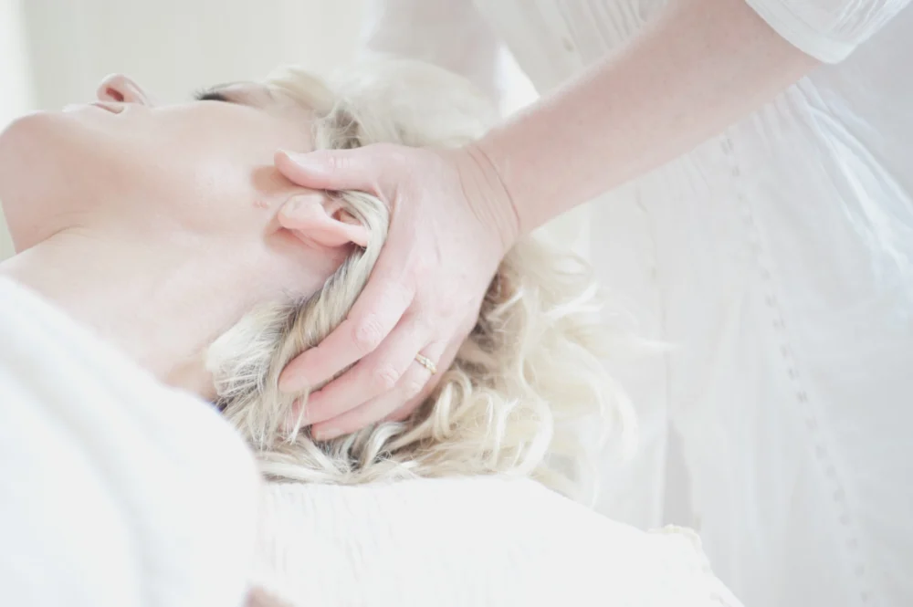
The Massage Paradox: Analysis of Trigger Factors
The specific instance of a patient feeling incapacitated after a face-down massage provides a clear example of how multiple factors converge to disrupt vestibular compensation. Analysts and physical therapists point to four primary triggers in this scenario:
- Visual Deprivation: In the face-down position (using a massage cradle), the patient’s vision is restricted to the floor or a small area. Since the brain relies heavily on vision to compensate for vestibular weakness, removing this "visual anchor" forces the brain to rely solely on the damaged vestibular nerve and the neck’s proprioceptors.
- Cervical Extension and Pressure: The positioning of the head in a massage cradle can put pressure on the upper cervical spine. If the patient has cervicogenic sensitivity, this pressure can distort the sensory input sent to the brain.
- Autonomic Shift: Massage induces deep relaxation, which shifts the body into a parasympathetic state. While generally positive, the sudden transition from deep relaxation to standing upright (orthostatic challenge) can be difficult for a compromised vestibular system to regulate.
- Fluid Dynamics: Prolonged periods of being prone can cause subtle shifts in the fluid pressure within the inner ear, which may affect those with a history of inflammation in the vestibular apparatus.
Official Responses and Clinical Recommendations
Leading vestibular specialists, including members of the Vestibular Disorders Association (VeDA), emphasize that recovery is not a straight line but a series of adaptations. Dr. Denise Schneider, a Doctor of Physical Therapy and vestibular specialist, notes that patient education is a critical component of treatment. The consensus among clinicians is that patients should not avoid activities like massage, but rather modify them to accommodate their system’s needs.
Clinical recommendations for patients with motion sensitivity include:
- Gradual Transitions: Taking five to ten minutes to sit up and acclimate after lying down before attempting to stand.
- Position Modification: Requesting a side-lying massage or using a reclined chair instead of a face-down table.
- Proprioceptive Grounding: Keeping a hand on a stable surface (like a wall or the massage table) when moving to provide the brain with a "grounding" signal.
- Targeted VRT: Engaging in exercises that specifically habituate the brain to the positions that trigger symptoms.
Broader Impact and Implications for Long-term Care
The long-term management of vestibular nerve injuries requires a shift in how the medical community views "recovery." A patient who is "mostly better" still requires an awareness of their environmental and physical triggers. The implications for the wellness industry are also significant; massage therapists and fitness instructors should be trained to recognize the signs of vestibular distress and offer modifications for clients with a history of balance disorders.
In conclusion, the vestibular nerve is a delicate and vital component of human functionality. Its injury marks the beginning of a lifelong process of neurological adaptation. While tight muscles, stress, and specific physical positions do not re-injure the nerve itself, they challenge the brain’s compensatory framework. Through a combination of physical therapy, stress management, and lifestyle adjustments, individuals can navigate these challenges, moving from a state of incapacitation to one of informed and proactive health management. The resilience of the human nervous system is remarkable, but it requires patience, understanding, and the right environmental supports to thrive after a vestibular disruption.
