
A Paradigm Shift in Early-Stage NPC Treatment
For decades, the recommended treatment for nasopharyngeal carcinoma (NPC), the most common epithelial malignancy of the head and neck, has been radiotherapy. This approach was largely dictated by the tumor’s inherent characteristics: a high likelihood of lymph node involvement and its secluded position within the nasopharynx, making complete surgical eradication notoriously difficult. This clinical reality has long positioned radiotherapy as the primary weapon, even for tumors confined to the nasal mucosal lining in their nascent stages, such as gastric cancer and colorectal neoplasia, where curative endoscopic surgery is well-established.
However, a confluence of factors is now prompting a re-evaluation of this entrenched approach. Enhanced health education initiatives and the widespread availability of early cancer screening programs have led to a substantial increase in the detection of Stage I NPC. Studies indicate that a significant proportion of newly diagnosed NPC patients, ranging from 47.1% to 51.1%, present with localized disease confined to the nasopharyngeal cavity. This burgeoning cohort of early-stage patients creates an urgent demand for therapeutic modalities that are less debilitating than conventional radiotherapy, thereby paving the way for the exploration of endoscopic surgical solutions.
Prior research has already laid a crucial foundation. Investigations into endoscopic nasopharyngectomy (ENPG) have demonstrated its feasibility for the dissection of newly diagnosed Stage I NPCs, and subsequent studies have helped delineate the specific patient populations who stand to benefit most from such procedures. A prospective clinical trial (NCT03353467) is currently underway, rigorously evaluating the efficacy and safety profile of endoscopic surgery for early-stage NPC, further underscoring the growing interest and investment in this therapeutic avenue.
Despite these advancements, existing ENPG techniques often present considerable technical hurdles. Their successful execution is heavily reliant on the surgeon’s intricate knowledge of nasopharyngeal anatomy and their advanced surgical dexterity, particularly when tumors involve critical areas like the pharyngeal recess and the lateral wall of the nasopharynx. To surmount these difficulties, a series of innovative surgical maneuvers and technologies have been developed. These include the "third-hand technique," the "cube-like resection" method, the "dumpling-making technique," and the utilization of a "pedicle nasal mucoperiosteal flap." These adaptations aim to facilitate en bloc resection (removal of the tumor in one piece), ensure the protection of the vital internal carotid artery (ICA), and effectively reconstruct the resultant surgical defects, all while operating within the confined spaces of the nasal and nasopharyngeal cavities.
Nevertheless, the pursuit of optimal surgical outcomes continues. The quest for a superior surgical perspective, a more refined understanding of surgical anatomy, and the standardization of surgical procedures remains paramount to achieving radical tumor removal while concurrently minimizing the risk of catastrophic ICA injury. It is within this context of ongoing innovation and persistent surgical challenge that the development of Nasopharyngeal Endoscopic Submucosal Dissection (NESD) emerges. This novel technique, honed over years of dedicated effort, is designed to enable the safe and complete resection of the entire nasopharyngeal mucosa, including the notoriously difficult-to-access pharyngeal recess and lateral wall, with an unprecedented level of internal carotid artery protection. This report marks the first formal documentation of the initial surgical experience utilizing the NESD technique for localized, early-stage NPC.
Case Study: A Pioneering Application of NESD
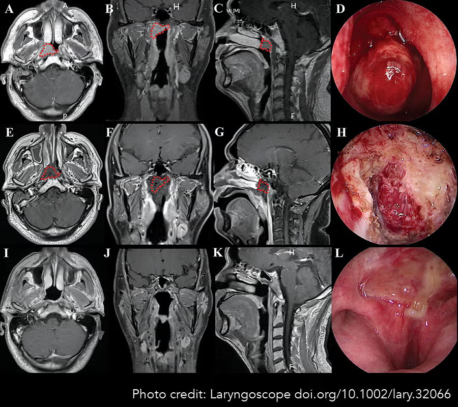
The genesis of this study involved a 40-year-old male patient who, during a routine health examination, was incidentally found to have a neoplasm on the right and posterior wall of his nasopharynx. Subsequent pathological examination confirmed the diagnosis of undifferentiated nonkeratinizing carcinoma. A comprehensive diagnostic workup, incorporating magnetic resonance imaging (MRI), positron emission tomography-computed tomography (PET-CT), and direct endoscopic evaluation, was undertaken to meticulously assess the extent of tumor invasion. Based on the eighth edition of the Union for International Cancer Control (UICC) staging system, the patient was definitively diagnosed with T1N0M0 Stage I NPC.
Crucially, after being presented with the treatment options, the patient opted for the NESD procedure, expressly declining radiotherapy. This decision highlights the growing patient preference for less invasive treatments when viable alternatives exist. The surgical plan meticulously defined the resection margins, encompassing the entire nasopharyngeal mucosa, guided by pre-operative MRI scans that clearly delineated the tumor’s boundaries. The research protocol received full approval from the clinical research ethics committee of the Fifth Affiliated Hospital of Guangzhou Medical University, and the patient provided comprehensive written informed consent, affirming his understanding and agreement to participate in this pioneering study.
The NESD Surgical Protocol: A Step-by-Step Innovation
The NESD procedure was performed under general anesthesia. Following standard pre-operative preparation and sterile draping of the head and neck region, the nasal mucosa was constricted using a solution of 1:10,000 epinephrine, administered under direct nasal endoscopic visualization. This step is critical for optimizing visualization and minimizing intraoperative bleeding.

Establishing the Surgical Field and Approach:
Imaging revealed the nasopharyngeal cavity to possess a roughly "cube-like" configuration, with the tumor situated on the right and posterior walls. The NESD was executed utilizing a 4-mm rigid endoscope, inserted via an oronasal approach. The surgical methodology comprised seven distinct phases, meticulously designed for the complete resection of the nasopharyngeal mucosa.
-
Resection Boundary Marking: The initial phase involved precisely marking the intended resection margins using electrocoagulation. This critical step ensures that the subsequent dissection adheres strictly to the pre-defined boundaries around the nasopharyngeal cavity, crucial for achieving adequate margins and preventing local recurrence.
-
Anterior Wall Mobilization: The anterior wall of the nasopharynx was carefully freed. This involved dividing the posterior mucosa of the nasal septum and polishing the vomer bone to effectively enlarge the operative space within the nasopharynx, thereby facilitating improved access for the endoscopic instruments.
-
Superior Wall Excision: The entire superior wall of the nasopharynx was then excised. This was achieved by dissecting the superior nasopharyngeal wall from the upper margin of the posterior nostril, following the bony surface all the way to the clivus. This maneuver creates a crucial opening and access point for subsequent steps.
-
Trans-Oral-Nasal Approach (TONA) Establishment: A key innovation in this technique is the establishment of a Trans-Oral-Nasal Approach (TONA). A mouth gag was employed to ensure the patient’s mouth remained open throughout the procedure. Two catheters were then introduced through the bilateral nasal cavities and the oropharyngeal cavity, with their tips subsequently retrieved from the oral cavity. These catheters served to suspend the soft palate, effectively retracting it from the surgical field. This combined TONA approach, integrating access from both the oral and nasal cavities, significantly broadens the surgical perspective, expands the operative workspace, and streamlines the surgical pathways, enhancing maneuverability and safety.
-
Posterior Wall Separation and Safety Tunnel Creation: The posterior wall of the nasopharynx was meticulously separated. Prior to this, a vital "safety tunnel" was established. This involved incising the mucosa of the posterior nasopharyngeal wall at the level of the cephalopharyngeus muscle and dissecting it along the surface of the longus capitis muscle up to the pharyngeal raphe. Paramount importance was placed on preserving the integrity of the longus capitis muscle membrane. Subsequently, the loose connective tissue within the submucosal plane, situated at the junction of the posterior and lateral nasopharyngeal mucosal walls, was carefully dissected. This dissection extended inferiorly to the apex of the petrous portion of the temporal bone and medially to the inner aspect of the internal carotid artery (ICA) sheath’s external orifice. This critical maneuver successfully established a safe tunnel that effectively separated the nasopharyngeal mucosa from the region of the ICA. The dissected connective tissue was then carefully removed inwards and upwards until it met the superior wall incision. This step is paramount for protecting the ICA from potential injury.
-
Left Lateral Wall Excision: The left lateral wall of the nasopharynx was then excised. The mucosa at the midpoint of the left Eustachian tube carina was incised, and the mucosal tissue was dissected along the submucosal space towards the parapharyngeal space. The pharynx was carefully recessed until the deepest point of the established safety tunnel was reached. The incisions were then meticulously converged with the superior and posterior wall incisions of the nasopharynx, allowing for the complete resection of the left nasopharyngeal wall. Throughout this process, extreme caution was exercised to ensure that the dissection did not extend beyond the boundaries of the safety tunnel, thereby safeguarding against injury to the parapharyngeal vessels and nerves.
-
Right Lateral Wall Excision: The right lateral wall was excised using an identical methodology to that employed for the left side. A right-sided safety tunnel was established following the same meticulous technique. The mucosa of the Eustachian tube carina was then divided and dissected until the right recess was reached via the submucosal space. Finally, this dissection converged with the contralateral nasopharyngeal superior and posterior wall incisions, following the surface of the longus capitis muscle. This comprehensive dissection ensured that the entire nasopharyngeal mucosa was completely detached from the tumor.
Specimen Retrieval and Wound Management:
The entire specimen, comprising the tumor encased within the whole nasopharyngeal mucosa, was retrieved orally in an en bloc fashion. Post-operative imaging confirmed complete tumor removal with adequate surgical margins. To prevent any risk of tumor implantation, the operative cavity was thoroughly irrigated with distilled water. Multiple biopsies were taken from the periphery of the incision margins for immediate pathological examination to confirm the absence of residual disease. A bioresorbable film was then applied to the nasopharyngeal wound to promote reepithelialization. A gelatin sponge and absorbable fibrin glue were used to pack the operative cavity, providing hemostasis and structural support. A final endoscopic assessment confirmed the absence of active bleeding from the oropharynx or bilateral nasal passages before the procedure was concluded.
Post-Operative Care and Follow-Up Protocols
Post-operative management was rigorous and systematic. Endoscopic evaluations of the wound healing process were conducted bi-weekly until complete reepithelialization was achieved. Subsequently, a structured follow-up schedule was implemented: assessments were planned every three months for the first three years, every six months from the fourth to the fifth year, and annually thereafter. Each follow-up included nasopharyngoscopy, head and neck MRI scans, and quantification of Epstein-Barr virus (EBV)-DNA copy numbers. Conventional examinations such as chest X-rays and abdominal ultrasounds were performed every six to twelve months, with more comprehensive whole-body imaging, including skeletal scintigraphy or PET/CT scans, utilized as clinically indicated.

Promising Initial Outcomes and Analysis
The initial NESD procedure for this Stage I NPC patient demonstrated highly encouraging results. The operation lasted 130 minutes, with a total blood loss of only 50 mL, underscoring the minimally invasive nature of the technique and the efficacy of intraoperative hemostasis. The patient reported no post-operative discomfort, such as headaches or nosebleeds.
Crucially, post-operative MRI scans, performed within one week of surgery, confirmed the complete removal of the primary tumor and the entire nasopharyngeal submucosa, with no evidence of residual disease. Pathological examination of the margin biopsies further validated the radical nature of the resection, reporting negative margins. The surgical wound healed completely within three weeks of the operation.
Over a comprehensive two-year follow-up period, the patient remained disease-free, exhibiting no signs of tumor recurrence or progression. Furthermore, the NESD technique proved to be exceptionally well-tolerated, with no sequelae such as xerostomia (dry mouth), hearing impairment, otitis media, trismus (difficulty opening the mouth), dysphagia (difficulty swallowing), or persistent nasal obstruction reported. These common debilitating side effects are often associated with conventional radiotherapy, making the absence of such complications a significant testament to the benefits of NESD.
Implications and Future Directions
The successful application of NESD in this initial case represents a significant advancement in the surgical management of early-stage nasopharyngeal carcinoma. The technique offers a compelling alternative to radiotherapy, achieving radical tumor resection with minimal invasiveness and a remarkably low morbidity profile. The ability to achieve complete mucosal resection, including challenging anatomical areas, while meticulously safeguarding vital structures like the internal carotid artery, addresses key limitations of previous endoscopic approaches.
The implications of this study are far-reaching. For patients diagnosed with early-stage NPC, NESD holds the promise of curative treatment with a significantly improved quality of life compared to radiotherapy. The reduced risk of long-term side effects associated with radiation therapy, such as xerostomia, radiation-induced fibrosis, and secondary malignancies, can profoundly impact a patient’s overall well-being and functional recovery.
The development of NESD also underscores the rapid progress in endoscopic surgical technology and surgical finesse. The meticulous planning, innovative instrumentation, and precise execution demonstrated in this case highlight the growing capabilities of minimally invasive surgery in treating complex oncological conditions.
However, it is important to acknowledge that this report details the initial experience with a single patient. While the outcomes are exceptionally promising, further research involving larger patient cohorts and longer follow-up periods is imperative to definitively establish the long-term efficacy, safety, and oncological control rates of NESD for Stage I NPC. Comparative studies directly contrasting NESD with current standard treatments, including radiotherapy and other endoscopic techniques, will be crucial for refining treatment algorithms and guiding clinical decision-making.
The ongoing prospective study (NCT03353467) will undoubtedly provide valuable data to further validate these findings. As the NESD technique continues to be refined and disseminated, it has the potential to redefine the standard of care for early-stage nasopharyngeal carcinoma, offering patients a less toxic and more effective path towards cancer remission and a higher quality of life. The commitment to surgical innovation, exemplified by the development of NESD, is a testament to the relentless pursuit of better outcomes for cancer patients worldwide.