Unveiling the Connection: A Large-Scale Investigation
The study, spearheaded by researchers including Laura A. Meliante, affiliated with Moorfields Eye Hospital and University College London, represents a significant stride in understanding the complex interplay between different sensory systems. Their meticulous analysis revealed that individuals demonstrating poorer hearing capabilities tended to exhibit slightly thinner inner and middle retinal layers. Crucially, this association remained statistically significant even after rigorous adjustment for a multitude of confounding factors, including age, sex, ethnicity, and various health and demographic indicators such as diabetes, smoking, alcohol consumption, body mass index, education level, socioeconomic deprivation, and even exposure to air pollution. This robust statistical control strengthens the credibility of the observed link, suggesting it is not merely a byproduct of general aging or lifestyle choices.
The sheer scale of the investigation, encompassing 32,613 adults with an average age of approximately 57 years, lends considerable weight to its conclusions. Of this substantial cohort, 5,475 participants, representing about 17% of the total, met the study’s predefined criteria for impaired hearing performance. This large sample size enhances the statistical power of the study, making it more likely to detect genuine, albeit subtle, associations that might be missed in smaller cohorts. Such a comprehensive approach is vital in identifying nuanced biological relationships within a diverse human population.
Precision in Measurement: Assessing Hearing and Retinal Structure
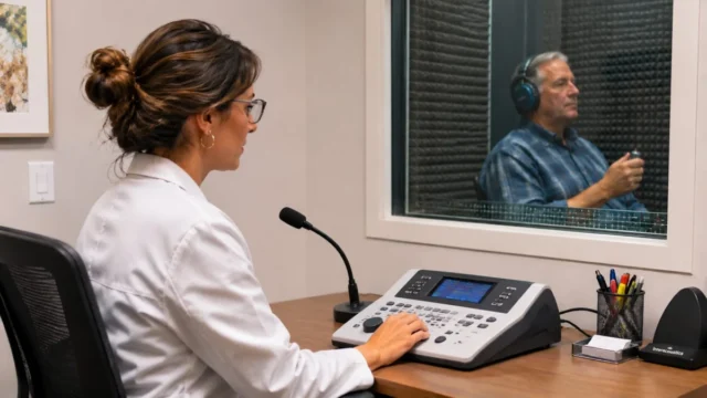
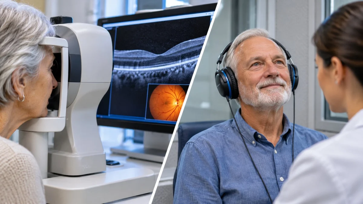
Unlike many previous studies that relied on subjective self-reports of hearing difficulties, the researchers employed a more objective and functionally relevant measure: speech reception thresholds obtained through speech-in-noise testing. This sophisticated audiological assessment evaluates an individual’s ability to discern speech amidst competing background noise – a critical component of real-world hearing ability and a common challenge for those with hearing impairment. By focusing on this functional aspect of hearing, the study provides insights that are directly pertinent to daily communicative experiences, which are often hampered more by noise than by quiet-environment hearing loss.
Retinal measurements were acquired using Optical Coherence Tomography (OCT), a non-invasive, high-resolution imaging technique that utilizes light waves to generate detailed cross-sectional images of biological tissues. OCT has revolutionized ophthalmology by enabling clinicians and researchers to visualize and quantify the individual layers of the retina with micron-level precision. This technology allows for the detailed examination of structures containing nerve cells, their intricate connections, and supporting tissues, providing a virtual histological view without the need for biopsy. The ability to differentiate and measure specific retinal layers, rather than just overall retinal thickness, was paramount to identifying the nuanced associations observed in this study, offering a level of anatomical specificity that previous methods could not achieve.
The core of the research involved a detailed statistical analysis to determine if variations in speech-in-noise performance correlated with specific changes in retinal thickness. The comprehensive adjustment for numerous potential confounders, as mentioned earlier, was vital to isolate the unique relationship between hearing and retinal structure from other influencing factors that might simultaneously affect both senses, thereby increasing the confidence in the observed association.
Dissecting the Findings: Specific Retinal Layers Affected
The study’s granular analysis pinpointed that poorer hearing performance was significantly associated with reduced thickness in several specific retinal structures, primarily within the inner and middle layers. These included the ganglion cell–inner plexiform layer (GCIPL) and the inner nuclear layer (INL). The GCIPL is particularly vital as it contains the cell bodies and dendrites of retinal ganglion cells, which are responsible for transmitting visual information from the eye to the brain via the optic nerve. Thinning in this layer could indicate subtle neuronal loss or altered connectivity, potentially impacting the efficiency of visual signal transmission. The INL, on the other hand, houses the cell bodies of various interneurons (bipolar, horizontal, and amacrine cells) that play crucial roles in processing and transmitting signals within the retina before they reach the ganglion cells, essentially acting as the retinal processing unit.
Furthermore, overall macular thickness also demonstrated an association with hearing performance. The macula is the central, most critical region of the retina, responsible for sharp, detailed central vision and color perception, essential for tasks like reading and recognizing faces. The findings indicated that for every 1-decibel increase in speech reception threshold (signifying poorer hearing performance), the total macular thickness was approximately 0.20 micrometers lower. While this difference appears miniscule – a micrometer is one-millionth of a meter – its statistical significance across a large population suggests a genuine biological correlation that warrants further investigation. The corresponding differences in individual retinal layers were, as expected, even smaller, underscoring the subtle nature of these changes.
When the researchers directly compared participants categorized as having impaired hearing against those with normal hearing performance, several retinal layers were indeed found to be significantly thinner in the impaired-hearing group. However, the association was not universal across all retinal structures. Notably, the study did not identify significant relationships involving the retinal nerve fiber layer (RNFL), the retinal pigment epithelium (RPE), or the photoreceptor layers. This specificity suggests that the link between hearing performance and retinal structure may involve particular neural or vascular tissues within the retina rather than a generalized, uniform thinning throughout the entire ocular structure. This nuanced observation provides crucial clues about the potential underlying biological mechanisms, guiding future research towards specific cellular and molecular pathways.
It is imperative to contextualize these findings: while statistically significant due to the large sample size, the observed differences in retinal thickness were quantitatively very small. Their direct clinical significance for individual patients, meaning whether these subtle changes translate into noticeable vision problems or serve as a standalone diagnostic marker, remains a subject for future investigation. This distinction between statistical significance and clinical relevance is a critical consideration in interpreting such research, emphasizing that these findings are currently more valuable for biological understanding than for immediate clinical application.
Exploring the Biological Nexus: Shared Vulnerabilities
The auditory and visual systems, though distinct in their sensory input and processing pathways, share fundamental biological underpinnings. Both rely on complex networks of specialized cells, intricate neural connections, and robust supporting biological processes, including vascular supply and metabolic regulation. The researchers propose that the observed associations could reflect shared neural vulnerability affecting both sensory systems. This "common cause" hypothesis suggests that a systemic factor, such as aging, inflammation, microvascular compromise, or neurodegeneration, might simultaneously impact the health and integrity of both the auditory and visual neural tissues.
This study adds to a burgeoning body of research exploring broader connections between sensory decline and systemic health. For instance, both hearing and vision impairment have independently been linked to an increased risk of cognitive decline and dementia. This has fueled significant interest in whether measurable changes in sensory organs could serve as early biomarkers or provide valuable insights into broader neurological health processes. The current study, while not directly investigating dementia or predicting cognitive decline, aligns with this growing paradigm, suggesting that the retina, often referred to as an "extension of the brain," might offer a window into wider neurological well-being. The implications of this are profound, as easily accessible retinal imaging could potentially offer insights into less accessible brain health.
The exact mechanisms linking hearing performance and retinal thickness remain undefined by this cross-sectional study. Potential explanations include shared genetic predispositions, common developmental pathways, microvascular dysfunction affecting both sensory neurovascular units, or shared inflammatory processes. For example, conditions like diabetes and hypertension are known to affect both retinal and inner ear microvasculature. While the study controlled for some of these systemic factors, residual confounding or more complex interactions cannot be entirely ruled out. Therefore, additional longitudinal and mechanistic research will be essential to unravel whether the findings reflect shared biological processes, common risk factors, or other intricate pathways. Understanding these pathways could open doors to preventative strategies that benefit multiple sensory systems.
The UK Biobank: A Foundation for Discovery
The UK Biobank stands as a monumental biomedical research resource, playing a pivotal role in facilitating studies of this magnitude and complexity. Launched in 2006, this prospective cohort study recruited 500,000 participants aged 40-69 years across the United Kingdom between 2006 and 2010. Its strength lies in its comprehensive data collection, which includes extensive genetic data, detailed lifestyle information, environmental exposures, biological samples (blood, urine, saliva), and sophisticated imaging data (brain, heart, body, eyes). Crucially, participants’ health is continuously tracked through linkage to their electronic health records, providing a dynamic longitudinal perspective on health and disease progression.
This unparalleled depth and breadth of data allowed Meliante and her team to conduct a highly robust analysis, controlling for an array of potential confounding variables that might obscure genuine biological associations. The inclusion of retinal OCT imaging data for a significant subset of participants, alongside objective hearing assessments, made this particular investigation feasible and highly informative. The UK Biobank serves as a testament to the power of large-scale population health research in uncovering subtle yet significant health insights that would be impossible to detect in smaller, less comprehensive studies, underscoring its role as a cornerstone for future biomedical breakthroughs.
Implications for Hearing Healthcare and Beyond
For audiologists, ophthalmologists, and other healthcare professionals involved in sensory health, these findings offer a fresh perspective on the potential interconnectedness of hearing and vision. It reinforces the notion that the human body’s systems are not isolated but rather intricately linked, suggesting a need for a more holistic approach to patient care. While not immediately altering clinical practice, the research encourages a broader consideration of a patient’s overall sensory health. An ophthalmologist might, for instance, consider asking about hearing difficulties, and an audiologist about vision concerns, leading to more integrated referrals.
The innovative use of OCT in this context is also noteworthy. While OCT is a cornerstone of ophthalmology, researchers are increasingly exploring its applications beyond the eye. For instance, as highlighted in the original context, researchers at the University of Southern California are actively developing an OCT-based cochleoscope. This ambitious project aims to enable real-time visualization of the delicate microstructures within the living inner ear – a challenging frontier in otology, given the ear’s intricate and often inaccessible anatomy. While the cochleoscope addresses a distinct clinical challenge (diagnosing inner ear pathologies), both endeavors underscore how advanced imaging technologies can help researchers gain unprecedented insights into sensory organ structures and their relationship to disease processes, potentially paving the way for novel diagnostic and therapeutic strategies that are minimally invasive and highly precise.
Looking Ahead: The Path to Clinical Utility
Despite the significant findings, the researchers prudently emphasize several limitations inherent in a cross-sectional study design. The analysis captured hearing performance and retinal measurements at a single point in time. Consequently, the findings cannot establish causality – it is unclear whether changes in hearing precede retinal thinning, whether retinal changes occur first, or if both are concurrently influenced by other deeper, underlying biological factors. Disentangling cause and effect will require prospective longitudinal studies that track individuals over extended periods, observing how these parameters evolve together. Such studies are costly and time-consuming but essential for moving beyond mere correlation.
Furthermore, the study utilized speech-in-noise performance as its primary measure of hearing, rather than a comprehensive audiologic evaluation that includes pure-tone audiometry across various frequencies. This specificity limits conclusions about particular types or degrees of hearing loss. While speech-in-noise testing is highly relevant for functional hearing, a full audiological profile would provide a more complete picture of auditory health, allowing for more granular associations to be identified.
Crucially, the study’s results do not, at this stage, establish retinal imaging as a screening or diagnostic tool for hearing loss. Leading experts in both ophthalmology and audiology would likely echo the researchers’ caution, stressing that the observed differences, though statistically significant, are too subtle for routine clinical application in diagnosing or monitoring hearing impairment in individual patients. Any potential clinical utility would require extensive validation through future research, demonstrating both high sensitivity and specificity.
The path forward involves robust longitudinal studies. These investigations will be instrumental in determining whether changes in hearing and retinal structure progress in parallel, whether one predicts the other, and ultimately, whether these relationships hold meaningful implications for clinical care, such as identifying individuals at risk for broader neurological decline or guiding integrated sensory interventions. For now, this research serves as a powerful testament to the interconnectedness of human biology, providing further compelling evidence that hearing and vision may be intricately linked through biological processes that extend far beyond the individual sensory organs. It unequivocally underscores the imperative for continued, rigorous scientific investigation before these fascinating findings can translate into actionable clinical guidelines or patient care strategies. The journey from correlation to causation, and eventually to clinical application, is often long and complex, but this study marks an important waypoint on that journey, inviting further exploration into the intricate tapestry of human sensory health.
Source: Meliante LA, Stuart KV, Meliante PG, et al. (2026). Association Between Retinal Layer Thickness and Hearing Performance in the UK Biobank: A Cross-Sectional Study. Investigative Ophthalmology & Visual Science, 67(13), 10. https://doi.org/10.1167/iovs.67.13.10.