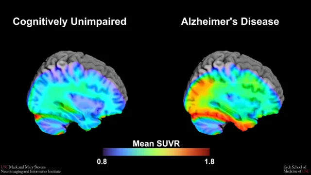
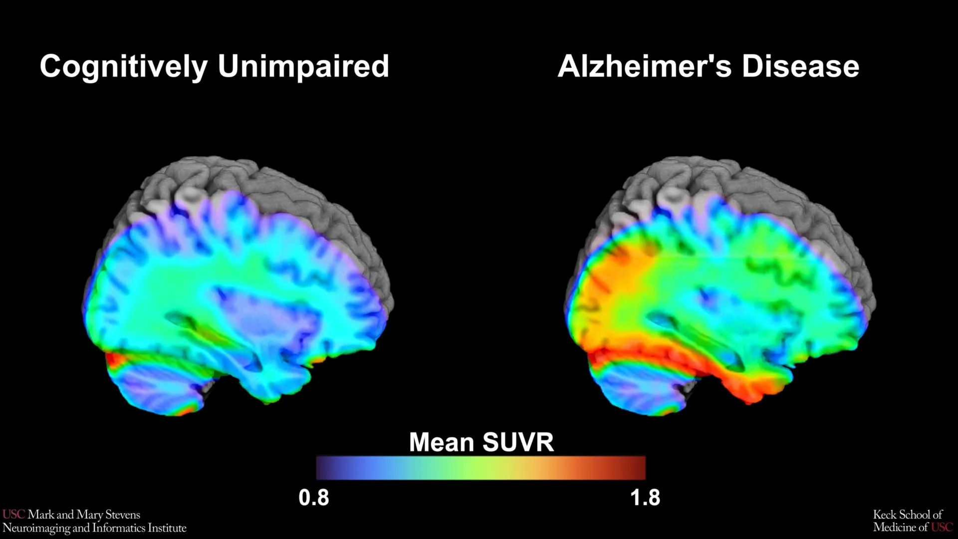
The research was conducted as part of the Health and Aging Brain Study-Health Disparities (HABS-HD), a comprehensive, multi-university collaboration led by the University of North Texas Health Science Center. Supported by the National Institute on Aging (NIA), the HABS-HD initiative represents one of the most ambitious efforts to date to understand how Alzheimer’s affects different ethnic and racial groups, moving beyond the historically homogenous data sets that have dominated neuroimaging research for decades.
The Science of Tau and the Evolution of Alzheimer’s Diagnostics
For much of the 20th century, an Alzheimer’s diagnosis was primarily clinical, based on observed cognitive decline and confirmed only through post-mortem examinations of brain tissue. The discovery of biomarkers—measurable biological indicators—transformed the field. Two proteins are central to this biological understanding: amyloid-beta, which forms plaques outside neurons, and tau, which creates neurofibrillary tangles inside neurons.
While amyloid plaques are often the first sign of the disease, appearing years or even decades before symptoms manifest, tau tangles are more closely correlated with the actual onset of memory loss and cognitive dysfunction. The ability to visualize these proteins in a living brain via Positron Emission Tomography (PET) scans has revolutionized early detection. The USC study utilized an advanced imaging tracer known as 18F-PI-2620. This second-generation tau PET tracer is designed to bind specifically to the tau aggregates associated with Alzheimer’s, providing a clearer and more precise signal than earlier technologies.
By analyzing the scans of over 675 older adults, the USC team sought to establish a "cut-point"—a mathematical threshold of tau density that indicates a transition from healthy aging to a disease state. Establishing such a benchmark is essential for clinical trials, as it allows researchers to identify participants who are in the early stages of the disease and most likely to benefit from experimental therapies.
Identifying the Medial Temporal Lobe as a Diagnostic Anchor
The study’s findings point to the medial temporal lobe (MTL) as the most reliable region for establishing this tau benchmark. Located deep within the brain, the MTL includes the hippocampus and the entorhinal cortex, areas fundamental to the formation and retrieval of memories. In Alzheimer’s disease, the MTL is typically one of the first regions to show significant tau accumulation.
The research team found that when tau levels in the MTL exceeded a specific threshold, there was a high probability of cognitive impairment. However, this correlation was not isolated. The data revealed that the tau cut-point was most effective at predicting cognitive decline when another biomarker, amyloid, was also present. This "double-hit" of amyloid and tau reinforces the prevailing "amyloid cascade hypothesis," which suggests that amyloid buildup triggers or accelerates the spread of tau, leading to neuronal death and cognitive failure.
"Establishing a cut-point in this region using 18F-PI-2620 marks an important step toward defining tau positivity for both research and clinical applications," said lead author Victoria R. Tennant, a PhD candidate in USC’s Neuroscience Graduate Program. The precision of the 18F-PI-2620 tracer allows for a more nuanced understanding of how tau accumulates in the MTL, potentially offering a more reliable diagnostic tool than previous methods.
Addressing the Diversity Gap in Neuroimaging
One of the most striking aspects of the study is its focus on health disparities. Historically, clinical trials for Alzheimer’s disease have been composed of up to 90% non-Hispanic White participants. This lack of diversity has led to diagnostic tools and treatments that may not be equally effective for all populations.
The USC study found that while the tau cut-point was highly effective for Hispanic and non-Hispanic White participants, it did not perform as expected for non-Hispanic Black participants. In this group, the established tau threshold did not reliably distinguish between those with cognitive impairment and those without. This suggests that the biological drivers of dementia in Black populations may involve different pathways, such as vascular issues, chronic inflammation, or other co-occurring pathologies that are not captured by tau PET scans alone.
Senior author Meredith N. Braskie, PhD, assistant professor of neurology at the Keck School of Medicine, emphasized the importance of this finding. "In non-Hispanic Black participants, the tau cut-point did not perform as expected. This suggests that other pathologies or conditions may be driving cognitive decline in this group. Our study is an important step toward better understanding how tau relates to cognition in diverse populations and has important implications for future clinical trials that aim to target tau."
Chronology and Context: The HABS-HD Initiative
The Health and Aging Brain Study-Health Disparities (HABS-HD) was launched to address the urgent need for inclusive Alzheimer’s research. Statistics show that Hispanic and Black Americans are disproportionately affected by Alzheimer’s and related dementias. Hispanic adults are approximately 1.5 times more likely, and Black adults are about twice as likely, to develop the disease compared to non-Hispanic White adults. Despite this, they remain underrepresented in the research that determines how the disease is diagnosed and treated.
The timeline of the current study reflects a multi-year effort to recruit and monitor a diverse cohort of participants. By utilizing a standardized protocol across multiple institutions, the HABS-HD project has built a robust database that includes blood-based biomarkers, genetic data, and advanced neuroimaging. The USC team’s analysis of the 675-person subset represents a critical milestone in this ongoing effort, providing the first major evidence of how tau PET benchmarks vary across ethnic lines.
Institutional Perspectives and Future Implications
The Mark and Mary Stevens Neuroimaging and Informatics Institute (Stevens INI) at USC has long been at the forefront of big-data approaches to brain health. Director Arthur W. Toga, PhD, noted that the findings are part of a broader shift toward personalized medicine.
"This type of imaging is critical for understanding who is at risk and how the disease develops," Toga stated. "These findings are just the latest to come from HABS-HD, which is the most comprehensive study of Alzheimer’s disease and related dementias in diverse communities. We hope this work will lead to more personalized care and better outcomes for all communities."
The implications of this research extend to the design of future clinical trials. If tau PET scans are used as a primary screening tool for drug trials, and if those scans are less reliable for Black patients, there is a risk that Black populations will continue to be excluded from or underserved by new treatments. The study highlights the need for a multi-modal approach to diagnosis—one that considers biological markers alongside social determinants of health, such as access to care, diet, and environmental factors.
Analysis: The Path Toward Precise Diagnostics
The identification of a tau cut-point in the medial temporal lobe provides a "biological ruler" that clinicians can use to measure the progression of Alzheimer’s. However, the study also serves as a cautionary tale against a "one-size-fits-all" diagnostic model. The failure of the tau benchmark in Black participants suggests that the medical community must look beyond the traditional amyloid/tau framework to fully understand dementia in diverse groups.
As the pharmaceutical industry continues to develop anti-amyloid and anti-tau therapies, such as the recently FDA-approved lecanemab and donanemab, the accuracy of these benchmarks becomes a matter of health equity. If the thresholds for "tau positivity" are based on data that does not accurately reflect the biology of all patients, the promise of these new drugs may remain out of reach for the very populations that suffer most from the disease.
The USC study, through its rigorous methodology and commitment to diversity, provides a roadmap for the future of Alzheimer’s research. It underscores that while technology like 18F-PI-2620 PET imaging offers unprecedented clarity, its interpretation must be grounded in an understanding of human diversity.
Research Support and Collaborative Effort
The study was a massive collaborative effort involving experts from the University of Southern California, the University of North Texas Health Science Center, the University of Wisconsin-Madison, Washington University in St. Louis, and the University of California, San Francisco.
The research was supported by several grants from the National Institute on Aging (NIA) and the National Institutes of Health (NIH), including R01AG054073 and U19AG078109. These grants are part of a federal push to increase the inclusion of underrepresented groups in aging research, a priority that has gained significant momentum over the last decade.
As the scientific community continues to parse the data from the HABS-HD study, the USC team’s findings will likely serve as a foundational reference point. By defining what "early Alzheimer’s" looks like in the brain across different populations, researchers are one step closer to a world where dementia can be detected, treated, and perhaps eventually prevented, for everyone.