The research, led by Seema Tiwari-Woodruff, a professor of biomedical sciences at the UC Riverside School of Medicine, suggests that the decline in motor function experienced by MS patients is not merely a result of external nerve damage but is driven by internal metabolic failure within the cells themselves. This discovery opens a new frontier for therapeutic intervention, shifting the focus from broad immune suppression to targeted mitochondrial protection.
The Pathological Framework: Inflammation and Energy Failure
Multiple sclerosis is traditionally characterized by two primary processes: inflammation and demyelination. The immune system mistakenly attacks the myelin sheath, the fatty, insulating layer that surrounds nerve fibers (axons). This insulation is vital for the rapid transmission of electrical impulses. When myelin is stripped away, signal conduction slows or fails entirely, leading to symptoms ranging from numbness and tingling to paralysis.
However, the UCR study posits that demyelination is only one part of a more complex degenerative cycle. Within the cerebellum, the loss of myelin appears to trigger a secondary, devastating effect on mitochondria. Mitochondria are organelles responsible for producing adenosine triphosphate (ATP), the chemical energy required for nearly every cellular function.
"Our study proposes that inflammation and demyelination in the cerebellum disrupt mitochondrial function, contributing to nerve damage and Purkinje cell loss," explained Professor Tiwari-Woodruff. The research team identified a significant reduction in the mitochondrial protein COXIV in demyelinated Purkinje cells. This protein is essential for the electron transport chain, the process by which mitochondria generate energy. When COXIV levels drop, the cell enters a state of energy crisis, eventually leading to programmed cell death.
The Critical Role of Purkinje Neurons in Human Mobility
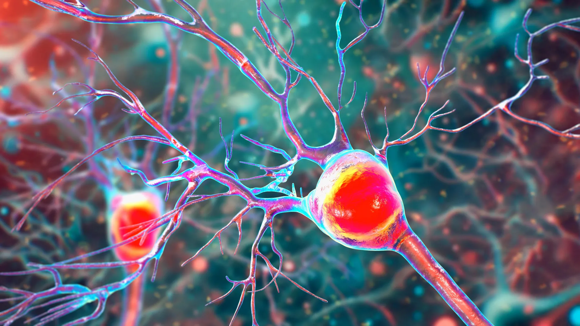
To understand the impact of this research, one must understand the function of Purkinje cells. These are among the largest and most complex neurons in the human brain, characterized by an extensive "tree" of dendrites that receive thousands of inputs from other parts of the nervous system. Located in the cerebellar cortex, they are the sole output for motor coordination from the cerebellar cortex to the rest of the brain.
Everyday activities—ranging from the fine motor skills required to type on a keyboard to the gross motor skills needed to maintain balance while walking—rely on the health of these cells. In MS, as Purkinje cells begin to malfunction and die, patients develop ataxia. Ataxia is a clinical term for a lack of voluntary coordination of muscle movements, which can include gait abnormality, speech changes, and difficulties with eye movement.
The UCR team analyzed postmortem cerebellar tissue from individuals who had been diagnosed with secondary progressive MS (SPMS). SPMS is a stage of the disease where disability worsens steadily, regardless of whether the patient experiences distinct "attacks" or relapses. The analysis revealed that the surviving Purkinje cells in these patients were severely compromised: they possessed fewer dendritic branches, showed significant myelin loss, and exhibited clear signs of mitochondrial failure.
Evidence from the EAE Mouse Model: A Chronological Analysis
To map the progression of this cellular decline, the researchers utilized the experimental autoimmune encephalomyelitis (EAE) mouse model. EAE is the standard laboratory model for studying MS, as it mimics many of the inflammatory and demyelinating features of the human disease.
By tracking the mice over time, the researchers established a clear chronology of neurodegeneration. They observed that myelin breakdown occurs early in the disease course, followed closely by the onset of mitochondrial dysfunction. Interestingly, while the "machinery" of the cells began to fail early on, the actual death of the Purkinje cells occurred later, as the disease transitioned into a more severe, chronic phase.
"The remaining neurons don’t work as well because their mitochondria, the energy-producing parts, start to fail," Tiwari-Woodruff noted. This finding is significant because it suggests a "window of opportunity" for medical intervention. If treatments can be administered after the initial inflammation but before the cells actually die, it may be possible to rescue the neurons and preserve motor function.
Supporting Data and Comparative Analysis
The study’s data was bolstered by a comparative analysis between healthy donor tissue and tissue from MS patients, provided by the National Institutes of Health’s NeuroBioBank and the Cleveland Clinic. The researchers focused on the expression of COXIV and other markers of mitochondrial health across different regions of the cerebellum.
The data showed a direct correlation between the degree of demyelination and the loss of mitochondrial proteins. In regions where myelin was still intact, Purkinje cells maintained relatively healthy mitochondrial profiles. However, in "lesioned" areas where myelin had been stripped away, the mitochondrial density was significantly lower. This suggests that the loss of the protective myelin sheath places an unsustainable metabolic demand on the axon, eventually exhausting the mitochondria.
This metabolic "burnout" is a leading theory in progressive MS. Without the insulation of myelin, the nerve fiber must expend significantly more energy to move ions across its membrane to propagate an electrical signal. If the mitochondria cannot keep up with this increased demand—or if they are damaged by the surrounding inflammation—the cell eventually runs out of fuel and undergoes apoptosis (cell death).
Implications for Future Therapeutic Strategies
Current treatments for MS, known as disease-modifying therapies (DMTs), are largely focused on the immune system. These drugs aim to reduce the frequency of relapses by preventing immune cells from entering the central nervous system and attacking myelin. While highly effective for relapsing-remitting MS (RRMS), these treatments have shown limited success in stopping the underlying neurodegeneration seen in progressive forms of the disease.
The findings from UC Riverside suggest that a shift in strategy is necessary. Rather than solely focusing on the immune response, future treatments may need to target neuroprotection and mitochondrial health.
Potential avenues for new therapies include:
- Mitochondrial Boosters: Compounds that enhance the efficiency of the electron transport chain or promote the biogenesis of new mitochondria.
- Antioxidant Support: Strategies to reduce the oxidative stress that damages mitochondria during periods of inflammation.
- Remyelination Therapies: Drugs that encourage the body to repair the myelin sheath, thereby reducing the metabolic load on the underlying neurons.
"Targeting mitochondrial health may represent a promising strategy to slow or prevent neurological decline and improve quality of life for people living with MS," Tiwari-Woodruff stated. This research aligns with a growing body of evidence suggesting that "energy failure" is a hallmark of many neurodegenerative diseases, including Parkinson’s and Alzheimer’s.
The Broader Scientific and Social Context
The research team is now expanding its scope to investigate whether mitochondrial damage affects other cell types in the cerebellum. This includes oligodendrocytes, the cells responsible for producing myelin, and astrocytes, which provide structural and metabolic support to neurons. Understanding how these cells interact during the disease process is crucial for developing a holistic treatment approach.
The study also highlights the critical importance of sustained funding for medical research. The National Multiple Sclerosis Society provided the funding for this project, but the researchers emphasized that public support and government investment are vital for turning laboratory discoveries into clinical realities.
"Cutting funding to science only slows progress when we need it most," Tiwari-Woodruff remarked, addressing the broader challenges facing the scientific community.
As the global population ages and the prevalence of neurological disorders rises, the economic and social burden of MS continues to grow. In the United States alone, the total economic impact of MS is estimated to be over $85 billion annually, including direct medical costs and lost productivity. Breakthroughs like the one at UC Riverside offer hope for reducing this burden by preserving the independence and mobility of those living with the disease.
Conclusion and Next Steps
The discovery that mitochondrial failure drives the loss of Purkinje cells provides a concrete target for the next generation of MS drugs. By identifying COXIV loss as a key marker of cellular decline, the UCR team has provided a roadmap for researchers to test new compounds that might stabilize these "powerhouses" before permanent damage occurs.
The study, authored by a team including Kelley Atkinson, Shane Desfor, Micah Feria, and others, stands as a testament to the power of combining human tissue analysis with animal models to solve complex biological puzzles. As the research continues, the focus will remain on translating these findings into therapies that can help MS patients maintain their balance, their movement, and their quality of life. For the 2.3 million people worldwide living with the uncertainty of MS, this shift toward mitochondrial-focused neuroprotection offers a significant and scientifically grounded path forward.