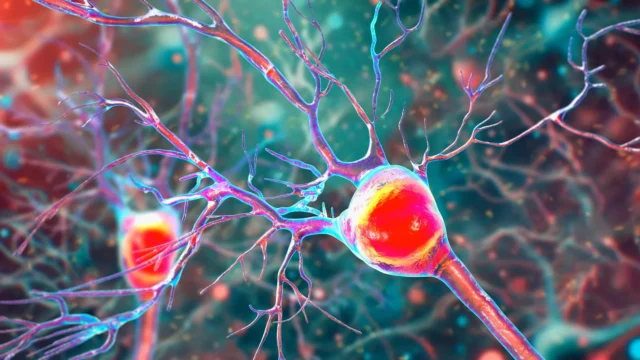

The research focuses on "enlarged perivascular spaces" (ePVS), which act as structural indicators of a compromised glymphatic system—the network responsible for clearing metabolic waste from the central nervous system. By identifying these anomalies during routine Magnetic Resonance Imaging (MRI) scans, clinicians may soon be able to flag Alzheimer’s risk years before memory loss or personality changes become debilitating. This discovery is particularly significant for the global medical community as it provides a non-invasive, accessible metric for a disease that has historically required expensive or invasive testing for definitive early-stage confirmation.
The Mechanics of Neurotoxic Waste Clearance
To understand the significance of the NTU Singapore findings, one must first examine the biological "plumbing" of the human brain. The brain is a highly metabolic organ that produces a significant amount of waste. Among the most dangerous of these byproducts are beta-amyloid and tau proteins. In a healthy brain, these proteins are flushed out through perivascular spaces—small, fluid-filled channels that surround the brain’s blood vessels.
When this drainage system functions efficiently, the brain maintains a delicate homeostatic balance. However, when these channels become "clogged" or less efficient, the fluid builds up, causing the perivascular spaces to enlarge. These enlarged spaces then become visible as distinct markers on MRI scans. For years, the presence of ePVS was noted by radiologists but its direct correlation with the specific pathology of Alzheimer’s disease remained a subject of debate. The NTU study provides the statistical evidence necessary to link these enlarged spaces directly to the buildup of toxic plaques and tangles that characterize Alzheimer’s.
Bridging the Research Gap in Asian Populations
One of the most critical aspects of this research is its focus on Asian demographics. For decades, the vast majority of Alzheimer’s clinical trials and longitudinal studies have been conducted on Caucasian populations in North America and Europe. This has created a significant data deficit, as neurodegenerative diseases do not manifest identically across all ethnic groups.
Associate Professor Nagaendran Kandiah, the study’s lead and Director of the Dementia Research Centre (Singapore) at NTU’s Lee Kong Chian School of Medicine (LKCMedicine), emphasized that genetic predispositions vary wildly by region. For instance, the apolipoprotein E4 (APOE4) gene is one of the most well-known genetic risk factors for Alzheimer’s. In Caucasian populations, this gene is present in approximately 50 to 60 percent of Alzheimer’s patients. However, in Singaporean patients, the prevalence of APOE4 is less than 20 percent.
This discrepancy suggests that other factors—potentially related to vascular health and environmental influences—play a more prominent role in the development of the disease among Asians. By examining a diverse cohort of nearly 1,000 Singaporeans of Chinese, Malay, and Indian descent, the NTU team has provided a more accurate profile of how Alzheimer’s progresses in a region that is home to a rapidly aging population.
Study Methodology and Findings
The research was a collaborative effort involving a multi-disciplinary team, including Justin Ong, a fifth-year medical student at LKCMedicine and the study’s first author. The team analyzed a total of 962 participants. This group was divided into two primary categories: those with normal cognitive function and those diagnosed with mild cognitive impairment (MCI). MCI is often considered a "prodromal" or precursor stage to full-blown dementia, characterized by noticeable but not yet disabling declines in memory and thinking speed.
The methodology involved a dual-track analysis:
- Neuroimaging: Participants underwent high-resolution MRI scans to identify and quantify the presence of enlarged perivascular spaces and white matter damage.
- Biochemical Analysis: Blood samples were taken to measure seven specific biomarkers associated with Alzheimer’s, including various forms of beta-amyloid and tau proteins, as well as markers for neuronal damage.
The results were definitive. Participants with mild cognitive impairment showed a significantly higher prevalence of enlarged perivascular spaces compared to the healthy control group. More importantly, the researchers found a direct correlation between these clogged drainage pathways and four of the seven blood-based biochemical markers of Alzheimer’s.
Comparing ePVS to White Matter Hyperintensities
Traditionally, clinicians have looked at white matter damage—often referred to as white matter hyperintensities—as a sign of "brain aging" or vascular disease. While the study confirmed that white matter damage is indeed linked to several Alzheimer’s markers, it also revealed a surprising nuance: in patients with early cognitive decline, the presence of enlarged perivascular spaces was a stronger predictor of Alzheimer’s-specific biochemical changes than white matter damage.
This finding suggests that ePVS is not just a secondary symptom of aging but a primary indicator of the early-stage Alzheimer’s process. Because ePVS is easily identifiable on routine MRIs already used to evaluate patients for headaches, dizziness, or minor cognitive complaints, it offers a "built-in" diagnostic tool that requires no additional specialized equipment or patient expenditure.
Expert Reactions and Clinical Implications
The medical community in Singapore and beyond has reacted with optimism to the findings. Dr. Rachel Cheong Chin Yee, a Senior Consultant at Khoo Teck Puat Hospital’s Department of Geriatric Medicine, noted the significance of the vascular connection. "These findings are significant because they suggest that brain scans showing enlarged perivascular spaces could potentially help identify people at higher risk of Alzheimer’s disease, even before symptoms appear," she stated.
Dr. Chong Yao Feng, a Consultant at the National University Hospital’s Division of Neurology, pointed out that the study challenges the traditional siloed view of brain diseases. Historically, "vascular dementia" (caused by blood vessel issues) and "Alzheimer’s disease" (caused by protein plaques) were treated as separate entities. Dr. Chong observed that this study demonstrates how the two conditions interact "in a synergistic manner." This means that a failure in the brain’s "plumbing" (vascular) directly accelerates the buildup of "trash" (Alzheimer’s proteins), creating a feedback loop of cognitive decline.
For general practitioners and neurologists, the study suggests a shift in how MRI reports are interpreted. If a patient presents with mild memory issues and their scan shows enlarged perivascular spaces, doctors should be more inclined to investigate for early-stage Alzheimer’s rather than dismissing the findings as general age-related changes.
Timeline and Future Trajectory of the Research
The NTU Singapore study is part of a broader, long-term effort to combat the rising tide of dementia in Asia. The timeline for this research began within the LKCMedicine Scholarly Project module, highlighting the institution’s commitment to involving medical students in high-impact research.
The research team is now moving into the longitudinal phase of the study. This involves tracking the original 1,000 participants over several years to observe how many of those with enlarged perivascular spaces eventually progress to a clinical diagnosis of Alzheimer’s dementia. This follow-up data will be crucial for establishing the "predictive power" of ePVS—determining exactly how many years in advance these brain blockages can predict the onset of symptoms.
Impact on Global Healthcare Strategy
As the world’s population ages, the economic and social burden of Alzheimer’s is expected to triple by 2050. Currently, the most accurate ways to detect the disease early are through Positron Emission Tomography (PET) scans—which are expensive and involve radiation—or lumbar punctures to collect cerebrospinal fluid, which are invasive and often avoided by patients.
The discovery that routine MRIs can provide similar early-warning signals could democratize Alzheimer’s screening. It allows for a "tiered" diagnostic approach:
- Screening: Identify ePVS on a standard MRI.
- Monitoring: Use blood tests to confirm the presence of amyloid and tau.
- Intervention: Start lifestyle changes, vascular health management, or newly approved anti-amyloid drug therapies before irreversible brain damage occurs.
By focusing on the brain’s waste removal system, the NTU Singapore team has not only found a new way to see the disease but has also highlighted a potential new target for treatment. If therapies can be developed to "unclog" these perivascular spaces or improve glymphatic flow, it may be possible to prevent the toxic buildup of proteins altogether, shifting the medical focus from managing dementia to preventing it.
This research serves as a vital reminder that local population studies are essential for global health. By looking specifically at the Asian brain, the scientists at NTU Singapore have provided a tool that will benefit clinicians and patients worldwide, ensuring that the next generation of Alzheimer’s care is both earlier and more inclusive.