
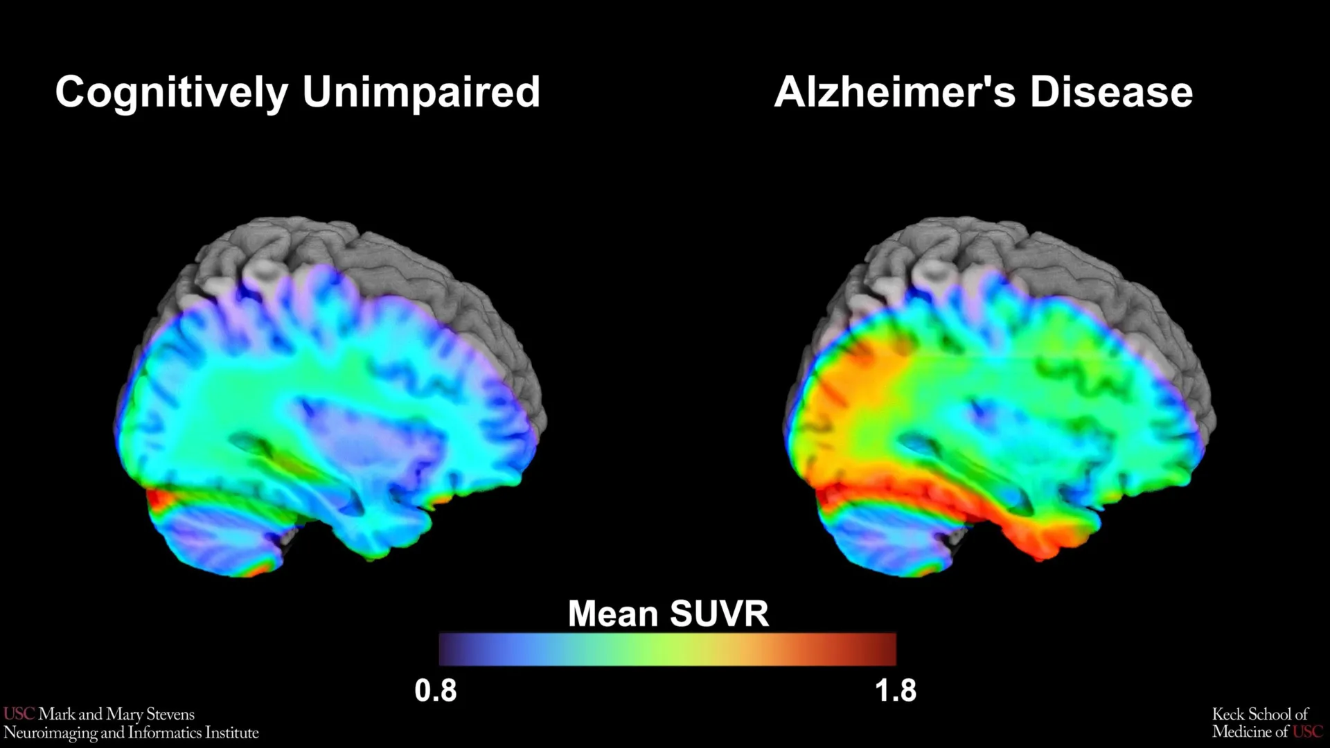
As the global medical community shifts toward precision medicine, the ability to accurately identify early-stage neurological decline has become paramount. The USC team’s research focuses on the "tau cut-point," a diagnostic threshold used to determine when the accumulation of tau protein in the brain reaches a level that suggests the early stages of Alzheimer’s disease or related cognitive disorders. By utilizing cutting-edge positron emission tomography (PET) technology, the researchers have provided a more nuanced understanding of how this protein behaves across different demographic groups, highlighting both the potential and the current limitations of standardized diagnostic markers.
The Biological Mechanism of Alzheimer’s Disease
To understand the significance of this new benchmark, it is essential to look at the underlying pathology of Alzheimer’s disease. For decades, the "amyloid cascade hypothesis" dominated the field, suggesting that the buildup of amyloid-beta plaques was the primary driver of the disease. However, while amyloid plaques appear early in the disease progression—often decades before symptoms emerge—they do not always correlate strongly with the severity of cognitive decline.
In contrast, tau protein, which forms "neurofibrillary tangles" within neurons, is more closely linked to the actual loss of memory and executive function. In a healthy brain, tau helps stabilize microtubules, the internal structures that transport nutrients within neurons. In Alzheimer’s, tau collapses into twisted strands, disrupting the transport system and eventually killing the cell. The progression of tau through the brain typically follows a predictable pattern, beginning in the medial temporal lobe—the region responsible for memory—before spreading to the cortex.
The USC study utilized an advanced imaging tracer known as 18F-PI-2620. This second-generation radioactive tracer is designed to bind specifically to tau aggregates with high affinity and low "off-target" binding, allowing for a much clearer visualization of protein density than previous technologies allowed.
Methodology and the Pursuit of the Tau Cut-Point
The research team analyzed data from over 675 older adults participating in the HABS-HD study. This cohort is unique in its diversity, specifically designed to address the historical underrepresentation of minority groups in Alzheimer’s research. The participants underwent comprehensive cognitive testing and advanced tau PET scans to establish a baseline for what constitutes "normal" aging versus "clinically relevant" protein accumulation.
The objective was to identify a "cut-point"—a mathematical threshold of tau density—that could distinguish between cognitively impaired individuals and those with normal age-related changes. By comparing the scans of those who showed impairment in cognitive tests with those who did not, the researchers sought a universal signal that could alert clinicians to the presence of AD pathology.
The team found that when tau levels in the medial temporal lobe exceeded a specific threshold, it served as a powerful indicator of cognitive impairment. However, the efficacy of this benchmark was found to be contingent on several biological and demographic factors, revealing a complex landscape of neurodegeneration that defies a one-size-fits-all approach.
Demographic Disparities in Diagnostic Accuracy
One of the study’s most striking findings was the variation in how the tau cut-point performed across different racial and ethnic groups. The benchmark proved highly effective for Hispanic and non-Hispanic White participants, particularly when another abnormal protein, amyloid, was also present. In these groups, the combination of high tau in the medial temporal lobe and the presence of amyloid plaques was a reliable predictor of Alzheimer’s-related cognitive decline.
However, the results for non-Hispanic Black participants were markedly different. In this group, the tau cut-point did not perform as expected, failing to reliably distinguish between those with and without cognitive impairment. This discrepancy suggests that the biological drivers of cognitive decline may differ across populations.
"In non-Hispanic Black participants, the tau cut-point did not perform as expected," noted senior author Meredith N. Braskie, PhD, assistant professor of neurology at the Keck School of Medicine. "This suggests that other pathologies or conditions may be driving cognitive decline in this group. Our study is an important step toward better understanding how tau relates to cognition in diverse populations and has important implications for future clinical trials that aim to target tau."
This finding underscores a growing concern in the medical community: diagnostic tools developed using data from predominantly White populations may not be universally applicable. Factors such as vascular health, chronic inflammation, and social determinants of health—including diet, environmental exposure, and lifelong stress—may play a larger role in cognitive decline for Black Americans, potentially overshadowing the role of tau protein as a primary biomarker.
The Role of the Medial Temporal Lobe
The focus on the medial temporal lobe is not incidental. This region, located deep within the brain, includes the hippocampus and is the primary hub for memory formation and spatial navigation. It is typically the first area to show tau accumulation in the earliest stages of Alzheimer’s, a phase often referred to as "prodromal" Alzheimer’s.
By establishing a cut-point specifically for the medial temporal lobe using the 18F-PI-2620 tracer, the USC researchers have provided clinicians with a specific "red zone" to monitor. Lead author Victoria R. Tennant, a PhD candidate in USC’s Neuroscience Graduate Program, emphasized that this is a major step toward defining "tau positivity"—a clinical designation that could determine whether a patient is eligible for certain treatments or clinical trials.
"While our findings support prior research linking medial temporal lobe tau to cognitive impairment, establishing a cut-point in this region marks an important step toward defining tau positivity for both research and clinical applications," Tennant stated. She also highlighted the need for more inclusive research, noting that the limited reliability of the tau marker in Black participants points to a need for studies that examine both biological and social determinants of the disease.
Chronology of the HABS-HD Initiative
The Health and Aging Brain Study-Health Disparities (HABS-HD) was launched to fill a critical gap in neurological science. Historically, over 90% of participants in Alzheimer’s clinical trials have been of European descent. This lack of diversity has led to a "knowledge gap" that complicates the treatment of Hispanic and Black populations, who are statistically more likely to develop Alzheimer’s than their White counterparts.
- Phase 1: Initial Recruitment: The HABS-HD project began by recruiting a large, diverse cohort in the Dallas-Fort Worth area, providing them with comprehensive medical exams, blood tests, and cognitive assessments.
- Phase 2: Introduction of Neuroimaging: As the study expanded, advanced neuroimaging, including MRI and PET scans, was integrated to correlate physical brain changes with cognitive performance.
- Phase 3: The Tau Study: The current research utilized the most recent advancements in PET tracer technology (18F-PI-2620) to focus specifically on the tau protein, leading to the identification of the new cut-points.
- Phase 4: Future Integration: The findings are now being integrated into larger datasets to help develop personalized risk profiles for patients based on their specific genetic and demographic backgrounds.
Broader Implications for Clinical Trials and Treatment
The implications of this research extend far beyond the laboratory. As pharmaceutical companies develop new "anti-tau" therapies, they require precise ways to identify which patients are most likely to benefit from these drugs. If a clinical trial uses a tau cut-point that is only accurate for certain populations, the resulting drug may be less effective—or even ineffective—for minority groups.
The USC study provides a roadmap for more inclusive trial designs. By recognizing that tau levels may mean different things in different populations, researchers can better tailor their inclusion criteria and ensure that new treatments are safe and effective for everyone.
Furthermore, the study highlights the necessity of a "multi-marker" approach to diagnosis. Because the tau cut-point was most effective when amyloid was also present, it suggests that clinicians should not rely on a single protein to make a diagnosis. Instead, a combination of amyloid PET, tau PET, and perhaps blood-based biomarkers will likely become the standard of care.
Expert Analysis and Future Directions
Arthur W. Toga, PhD, director of the Stevens INI, emphasized that the HABS-HD study is the most comprehensive effort to date to understand Alzheimer’s within diverse communities. "This type of imaging is critical for understanding who is at risk and how the disease develops," Toga said. "HABS-HD has already produced key findings related to ethnic variations in AD biomarkers, the influences of social determinants on cognitive health, and vascular contributions to dementia."
The goal, according to Toga and his colleagues, is to move toward a future of "personalized neuro-care." In this future, a patient’s risk for Alzheimer’s would be assessed not just by their age or memory scores, but by a sophisticated analysis of their brain imaging, genetic markers, and life history.
The USC study serves as a call to action for the scientific community to continue investigating why tau protein behaves differently across demographics. Potential avenues for future research include investigating the role of "co-pathologies," such as TDP-43 (another protein associated with dementia) or small vessel disease, which may be more prevalent in certain groups and could interact with tau in ways that are not yet fully understood.
As the global population ages, the burden of Alzheimer’s disease is expected to grow. By refining diagnostic benchmarks like the tau cut-point and ensuring they are validated across all populations, researchers at USC and their partners are ensuring that the next generation of Alzheimer’s care is more accurate, more inclusive, and ultimately more effective for all.