
A groundbreaking three-year research initiative, spearheaded by Dr. Yingying Wang at the University of Nebraska–Lincoln, has delved into the complex interplay between brain activity, sensory integration, and speech perception outcomes in individuals utilizing cochlear implants (CIs). The study, a collaborative effort involving the University of Nebraska Medical Center (UNMC) and Ohio State University, represents a significant step forward in understanding how neuroimaging techniques could potentially predict which candidates are most likely to achieve optimal benefit from cochlear implantation, thereby revolutionizing patient selection and personalized rehabilitative strategies.
The Quest for Predictable Outcomes in Cochlear Implantation
For individuals grappling with severe to profound hearing loss, cochlear implants stand as a transformative technological marvel, offering unparalleled access to sound and dramatically improving communication abilities and overall quality of life. Unlike conventional hearing aids, which merely amplify residual hearing, CIs are sophisticated electronic devices designed to bypass damaged portions of the inner ear. They directly stimulate the auditory nerve, transmitting electrical signals to the brain which are then interpreted as sound. This intricate process can restore a sense of hearing that profoundly impacts daily life, from engaging in conversations to appreciating music and environmental sounds.
However, despite their profound potential, the efficacy of cochlear implants is not uniform. Outcomes vary widely among recipients, presenting a persistent clinical challenge: understanding the factors that predispose certain patients to greater success remains an important, unanswered question for audiologists and otolaryngologists globally. This variability underscores the critical need for advanced predictive tools that can guide clinicians in patient counseling, managing expectations, and tailoring post-implantation rehabilitation.
Unpacking the Technology: How Cochlear Implants Work
To fully appreciate the significance of Dr. Wang’s research, it is essential to understand the fundamental mechanics of cochlear implants. A CI system comprises two main components: an external processor and an internal implant. The external sound processor, worn behind the ear, captures sound waves and converts them into digital information. This information is then transmitted wirelessly to the internal implant, surgically placed under the skin behind the ear. The internal implant features a receiver/stimulator that decodes the digital signals and sends them to an electrode array. This array is carefully threaded into the cochlea, the snail-shaped, fluid-filled structure of the inner ear. The electrodes then directly stimulate the auditory nerve fibers, bypassing the damaged hair cells that are typically responsible for converting sound vibrations into neural signals. These electrical impulses are then sent to the brain, where they are interpreted as sound.
The success of this complex neural pathway hinges not only on the integrity of the auditory nerve but also, crucially, on how well the brain’s intricate networks process and adapt to these novel electrical signals. For individuals who have experienced prolonged periods of profound hearing loss, the auditory cortex—the part of the brain responsible for processing sound—may have undergone significant changes, a phenomenon known as neuroplasticity. This adaptability of the brain, while sometimes beneficial, can also present hurdles, as the brain must re-learn to interpret these new, electrically generated sound representations.
A Deeper Dive into the Brain’s Role: The University of Nebraska–Lincoln Study
Dr. Yingying Wang, an associate professor of special education and communication disorders at the University of Nebraska–Lincoln, leads the Neuroimaging for Language, Literacy and Learning Lab and serves as resident faculty in the Center for Brain, Biology and Behavior. Her affiliations also extend to the Nebraska Center for Research on Children, Youth, Families and Schools, underscoring the multidisciplinary nature of her work. Her recent National Institute on Deafness and Other Communication Disorders (NIDCD)-funded project aimed to unravel the neurological underpinnings of varying CI outcomes.
The research team, which included co-principal investigator Michelle Hughes, professor of special education and communication disorders; Jonathan Hatch, an otolaryngologist and assistant professor at UNMC who performed the cochlear implant procedures; and Hongying (Daisey) Dai, an associate professor at UNMC serving as the project’s statistician, employed advanced neuroimaging techniques to observe brain activity. The central hypothesis was that individual differences in brain network function and sensory integration capabilities could predict how effectively a CI user would perceive speech, especially in challenging listening environments.
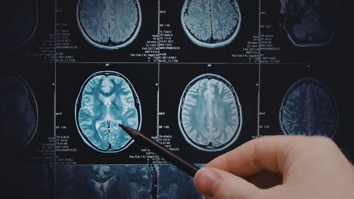
Neuroimaging Techniques: Peering into the Brain
The study harnessed the power of two prominent neuroimaging tools:
-
Resting-state Functional Magnetic Resonance Imaging (fMRI): This technique measures brain activity by detecting changes in blood flow. When a particular area of the brain is more active, it requires more oxygen, leading to an increase in blood flow to that region. Resting-state fMRI specifically assesses brain connectivity by examining spontaneous fluctuations in blood oxygenation levels when a person is not performing a specific task. This allows researchers to map out functional networks and understand how different brain regions communicate with each other. In the context of CI users, it can reveal how auditory pathways and related networks are organized and function, both before and after implantation.
-
Functional Near-Infrared Spectroscopy (fNIRS): A non-invasive optical imaging technique, fNIRS measures changes in hemoglobin concentrations associated with neural activity. It uses near-infrared light to penetrate the scalp and skull, allowing researchers to monitor brain activity in cortical regions. fNIRS is particularly advantageous for studies involving movement or in populations where traditional fMRI might be challenging, such as young children or individuals who find lying still difficult. For this study, fNIRS provided crucial insights into how the brain processes sound, especially when examining sensory integration.
Through these techniques, the research team examined various factors, including the recipient’s age at implantation and pre-existing hearing thresholds, and their influence on the brain’s connectivity and network efficiency. A particularly novel aspect of the investigation was the exploration of a measure termed the visual analog of temporal envelope. This measure reflects the brain’s ability to integrate sensory information across different modalities, specifically how visual cues might influence auditory processing. The researchers observed that this visual analog appeared to bolster speech perception for cochlear implant users, particularly in moderately noisy environments, suggesting a crucial role for cross-modal sensory integration in optimizing CI performance. This finding aligns with growing evidence that the brain doesn’t process sensory information in isolation but rather integrates cues from multiple senses to form a coherent perception of the world.
A Chronology of Brain Adaptation: Before and After Implantation

A key strength of the study design was its longitudinal approach, tracking brain changes in cochlear implant candidates both before and after surgery. This "before and after" methodology is critical for understanding the dynamic processes of neural adaptation.
-
Pre-implantation Assessment: Prior to receiving their cochlear implants, participants underwent extensive neuroimaging. The primary objectives were to identify brain regions that demonstrated responsiveness to speech sounds and to confirm the anatomical integrity of the auditory nerve. This baseline data was essential for understanding the pre-existing neural landscape upon which the CI would impose new auditory input. It also helped to rule out any underlying neurological conditions that might impact CI success.
-
Post-implantation Monitoring: Following the surgical procedure, the research team meticulously monitored changes in brain activity and speech perception during a series of follow-up visits. This phase was crucial for observing how the brain began to interpret the new electrical signals from the implant and how this adaptation correlated with improvements in speech understanding. The goal was to gain a deeper understanding of why some CI recipients achieve robust speech perception outcomes, while others struggle despite having functionally intact devices.
Dr. Wang eloquently likened this intricate adaptation process to adjusting to a new pair of eyeglasses, emphasizing that the brain must actively learn to interpret and make sense of this entirely novel form of sensory input. She elaborated on the profound neuroplasticity involved: "For people who have been deaf for years, this region [the auditory cortex] has often been recruited for vision or touch. Now sound signals are rushing in again, and the brain needs to relearn how to process them." This statement highlights the significant reorganization that can occur in the brain in response to sensory deprivation and the subsequent challenge and opportunity presented by cochlear implantation. The brain, being remarkably adaptable, must re-allocate neural resources and establish new pathways to effectively process the incoming auditory information.
Challenges in Research and the Path Forward
Despite the significant insights gleaned, the study encountered a common challenge in clinical research: patient recruitment. While twelve adults with severe-to-profound hearing loss completed comprehensive pre-surgical evaluations, only five participants were able to complete the post-surgical assessments. This limited sample size, while yielding valuable pilot data, restricts the generalizability of the findings and the statistical power to draw definitive conclusions.
Recognizing the immense potential of this preliminary work, Dr. Wang is actively pursuing additional federal funding to expand the research. The proposed follow-up study aims to address the sample size limitation by recruiting a larger cohort of cochlear implant candidates in Columbus, Ohio, alongside a continued focus on comparison participants at the Nebraska research site. This multi-site approach would significantly enhance the study’s power and scope, allowing for more robust statistical analysis and a broader understanding of the factors influencing CI outcomes across diverse populations.
Potential for Future Interventions and Clinical Impact
The implications of this research extend far beyond mere prediction; they open avenues for proactive intervention. Looking ahead, Dr. Wang envisions a future where pre-surgical interventions could be developed to prime the brain for optimal adaptation to auditory input from a cochlear implant. "By examining the brain’s neuroplasticity, we may be able to determine pre-surgery interventions to benefit patients," Wang stated. Such interventions might include targeted auditory training, cognitive exercises, or even pharmaceutical approaches designed to enhance neural plasticity and facilitate the brain’s re-learning process.
The ability to accurately predict patient outcomes before surgery would empower clinicians to provide more personalized counseling, set realistic expectations, and tailor rehabilitation programs to individual needs. For instance, if neuroimaging indicated a patient might face greater challenges in adapting, specific pre-surgical therapies or intensified post-surgical training could be recommended. Conversely, patients predicted to achieve excellent outcomes could proceed with greater confidence. This would represent a paradigm shift in audiological care, moving from a reactive approach to a proactive, precision medicine model.
Ultimately, a deeper understanding of how the brain adapts to cochlear implant stimulation could lead to:
- Improved Patient Selection: More precise criteria for identifying candidates most likely to benefit.
- Personalized Rehabilitation: Development of individualized therapy plans based on neurological profiles.
- Enhanced Outcomes: Strategies to maximize speech perception and quality of life for all CI recipients.
- Reduced Variability: Narrowing the gap between the best and worst outcomes, ensuring more consistent success.
This research, supported by robust data and meticulous methodology, is a testament to the power of interdisciplinary collaboration in tackling complex medical challenges. The findings contribute significantly to the fields of audiology, neurosciences, and rehabilitation, promising a future where cochlear implant recipients can embark on their journey to hearing with a clearer roadmap to success. As the scientific community continues to unravel the mysteries of brain function and sensory processing, studies like Dr. Wang’s bring us closer to realizing the full potential of life-changing technologies like cochlear implants, ensuring that more individuals can experience the richness of sound and connection.
References
Ellsworth, C. T., et al. (2025). Age-Related Hearing Decline and Resting-State Networks. American Journal of Audiology. DOI: 10.1044/2025_aja-25-00025.
Gao, Z., et al. (2025). Visual Analog of Temporal Envelope Benefits Speech Processing in Cochlear Implant Users: A Pilot Functional Near-Infrared Spectroscopy Study on the Associations Between Audiovisual Benefit, Listening Environment, and Peripheral Neural Health. Ear & Hearing. DOI: 10.1097/aud.0000000000001709.
Source: UNL, AJA, E&H