
For individuals grappling with severe to profound hearing loss, cochlear implants (CIs) represent a transformative technology, offering a pathway to sound that can profoundly enhance communication abilities and overall quality of life. However, despite their undeniable success for many, the effectiveness of CIs varies significantly among recipients, leaving clinicians and patients alike with a critical unanswered question: which patients are most likely to achieve optimal outcomes? A landmark three-year research project led by Dr. Yingying Wang at the University of Nebraska–Lincoln has delved into this complex issue, meticulously examining how brain activity and sensory integration mechanisms may fundamentally influence speech perception outcomes in CI users. This ambitious study has explored the innovative potential of neuroimaging techniques to predict which candidates might experience the most substantial benefits from implantation, paving the way for a more personalized and effective approach to cochlear implantation.
Understanding Cochlear Implants: A Technological Marvel Against Hearing Loss
Hearing loss is a global health challenge, affecting hundreds of millions worldwide. According to the World Health Organization (WHO), over 5% of the world’s population – or 430 million people – require rehabilitation for disabling hearing loss. A significant portion of these individuals experience severe to profound hearing loss, where traditional hearing aids, which merely amplify sound, often prove insufficient. This is where cochlear implants enter the picture as a sophisticated medical intervention. Unlike hearing aids, CIs are complex electronic devices designed to bypass damaged portions of the inner ear, specifically the cochlea, and directly stimulate the auditory nerve. This direct stimulation provides a representation of sound signals to the brain, effectively restoring a sense of hearing.
The history of cochlear implants is a testament to scientific ingenuity and perseverance. Early experimental devices emerged in the 1950s and 60s, with significant breakthroughs in the 1970s and 80s leading to the first multi-channel implants. The U.S. Food and Drug Administration (FDA) approved the first cochlear implant for adults in 1984, followed by approval for children in 1990. Since then, technological advancements have continually refined CI design, improving sound processing, speech understanding, and noise reduction capabilities. Today, CIs are a standard treatment for eligible individuals with severe to profound sensorineural hearing loss. However, the degree of benefit, particularly in complex listening environments, remains highly variable. Factors such as the duration of hearing loss, age at implantation, health of the auditory nerve, and crucially, the brain’s ability to process and interpret the new electrical signals, all play a role in determining individual outcomes. The effectiveness of these devices, therefore, depends not only on the integrity of the auditory nerve but also significantly on how well the associated brain networks function and adapt.
The Nebraska-Led Investigation: A Deep Dive into Brain’s Adaptability
Dr. Yingying Wang, a distinguished associate professor of special education and communication disorders, spearheads the Neuroimaging for Language, Literacy and Learning Lab and serves as resident faculty in the Center for Brain, Biology and Behavior. She is also affiliated with the Nebraska Center for Research on Children, Youth, Families and Schools. Her research team embarked on this critical inquiry through a collaborative effort involving experts from the University of Nebraska–Lincoln, the University of Nebraska Medical Center (UNMC), and Ohio State University. This interdisciplinary approach, leveraging diverse expertise in audiology, neuroimaging, otolaryngology, and statistics, underscores the multifaceted nature of the challenge. The study, funded by the National Institute on Deafness and Other Communication Disorders (NIDCD), highlights the national importance placed on understanding and improving outcomes for individuals with hearing loss.
The core objective of Wang’s investigation was to move beyond traditional audiological assessments and explore the underlying neural mechanisms that dictate CI success. The research team meticulously examined how various factors, including the patient’s age at implantation and their pre-existing hearing thresholds, influenced the brain’s structural and functional characteristics, specifically its connectivity and network efficiency. Brain connectivity refers to the intricate web of pathways through which different brain regions communicate, while network efficiency relates to how quickly and effectively information can be processed and transmitted across these networks. A key innovation of the study was the investigation into the role of a measure known as the visual analog of temporal envelope. This intriguing measure reflects the brain’s capacity for sensory integration – the ability to combine information from different senses, in this case, visual cues with auditory input from the CI. According to the researchers, this measure demonstrated a promising correlation, appearing to significantly support speech perception for cochlear implant users, particularly in challenging moderately noisy environments where distinguishing speech from background sound is often difficult.
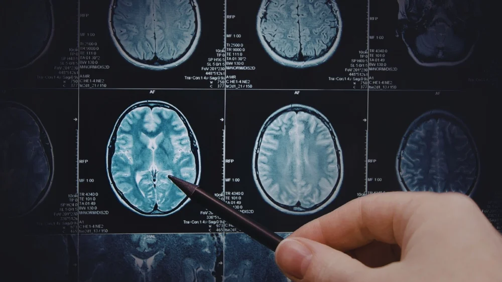
Neuroimaging: Illuminating the Brain’s Response to Sound
The study made extensive use of advanced neuroimaging tools, demonstrating their potential value in unraveling the intricate ways the brain processes sound following implantation. Specifically, the team employed resting-state functional magnetic resonance imaging (fMRI) and functional near-infrared spectroscopy (fNIRS).
Resting-state fMRI is a non-invasive technique that measures brain activity by detecting changes in blood flow. It allows researchers to map brain networks and assess their connectivity when an individual is not engaged in a specific task, providing insights into the brain’s intrinsic organization. This was crucial for understanding baseline brain function and how it might reorganize after CI activation. Functional near-infrared spectroscopy (fNIRS), another non-invasive neuroimaging technique, measures changes in blood oxygenation and volume in the cerebral cortex. It offers a more portable and often more comfortable alternative to fMRI, particularly useful for studying populations where fMRI might be challenging. Both techniques provide critical data on neural activity, offering a window into how the brain adapts to and integrates the novel auditory input from a cochlear implant. These tools were instrumental in tracking dynamic changes within the brain, offering a deeper understanding of the neurophysiological underpinnings of CI success.

Tracking Brain Changes: A Longitudinal Perspective
A cornerstone of Wang’s project was its longitudinal design, which allowed researchers to track brain activity in cochlear implant candidates both before and after surgery. This pre- and post-implantation comparison is vital for understanding the adaptive processes of the brain. Prior to implantation, participants underwent rigorous neuroimaging to identify specific brain regions that were responsive to speech sounds and to confirm the integrity of the auditory nerve. An intact auditory nerve is a fundamental prerequisite for CI success, as it is the direct pathway for electrical signals to reach the brain.
Following surgery, the research team continued to monitor changes in brain activity and speech perception during carefully scheduled follow-up visits. This systematic tracking aimed to uncover the neural correlates of improved speech understanding and, critically, to better understand the reasons behind the observed variability in outcomes among CI recipients. Dr. Wang eloquently compared the brain’s adaptation process to adjusting to a new pair of eyeglasses. Just as new glasses require the visual system to recalibrate and interpret a slightly different visual input, the brain, upon receiving signals from a CI, must learn to interpret this entirely new form of sensory information. She noted, “For people who have been deaf for years, this region has often been recruited for vision or touch. Now sound signals are rushing in again, and the brain needs to relearn how to process them.” This statement underscores the remarkable neuroplasticity of the human brain – its ability to reorganize itself by forming new neural connections and pathways throughout life, particularly in response to sensory experiences or injury. In individuals with prolonged hearing loss, the auditory cortex may be repurposed for processing visual or tactile information, and a successful CI outcome hinges on the brain’s capacity to re-engage these areas for auditory processing.
Challenges, Collaboration, and Future Horizons
The execution of such pioneering research is not without its obstacles. Recruitment, a common challenge in specialized clinical studies, proved particularly difficult for this project. While twelve adults with severe-to-profound hearing loss completed comprehensive pre-surgical evaluations, only five participants were able to complete the subsequent post-surgical assessments. This limited sample size, though providing valuable pilot data, restricts the generalizability of the findings and underscores the need for larger cohorts. Despite this hurdle, the initial findings are robust enough to warrant further investigation.
Recognizing the immense potential of this preliminary work, Dr. Wang is actively seeking additional federal funding to expand the research. The proposed follow-up study envisions a broader recruitment strategy, including additional cochlear implant candidates from a new research site in Columbus, Ohio. Concurrently, the Nebraska research site would focus on comparison participants, providing a crucial control group for a more comprehensive analysis. This expansion is critical to validating the initial findings, refining the neuroimaging predictors, and ultimately developing robust clinical tools.
The research team behind this significant endeavor included Dr. Michelle Hughes, professor of special education and communication disorders and co-principal investigator; Dr. Jonathan Hatch, an otolaryngologist and assistant professor at UNMC who performed the cochlear implant procedures; and Dr. Hongying (Daisey) Dai, an associate professor at UNMC who served as the project’s statistician, providing essential analytical expertise. This collaborative strength across diverse medical and scientific disciplines was fundamental to the study’s design and execution.
Implications for Personalized Medicine and Pre-Surgical Interventions
Looking ahead, the implications of Dr. Wang’s research are profound, holding the promise of revolutionizing how cochlear implant candidates are evaluated and managed. A primary aspiration is that further research will help identify pre-surgical interventions that could proactively improve cochlear implant outcomes. By understanding the brain’s neuroplasticity – its inherent capacity for change and adaptation – clinicians may be able to prescribe targeted therapies or training regimens even before surgery. “By examining the brain’s neuroplasticity, we may be able to determine pre-surgery interventions to benefit patients,” Wang emphasized.
Imagine a future where a patient, identified through neuroimaging as having a brain that might struggle with adaptation, could undergo a personalized regimen of auditory training or cognitive exercises before their CI surgery. Such interventions could prime the brain, making it more receptive to the new auditory input and accelerating the learning process post-implantation. This proactive approach could significantly enhance speech perception outcomes, reduce rehabilitation time, and maximize the overall benefit derived from the implant. For instance, pre-surgical auditory training could involve exercises designed to reactivate dormant auditory pathways or strengthen neural connections related to sound processing, thereby setting the stage for more effective post-surgical adaptation.
Beyond pre-surgical preparation, the ability to predict outcomes more accurately would enable clinicians to provide more realistic expectations to patients and their families. This personalized counseling, based on objective neural markers, could empower individuals to make more informed decisions about their treatment options. Furthermore, understanding how the brain adapts to cochlear implant stimulation could ultimately help clinicians develop tailored post-surgical rehabilitation strategies, optimizing therapy to individual neurological profiles rather than relying on a one-size-fits-all approach. This shift towards personalized medicine in audiology represents a significant step forward, promising a future where every CI recipient has the best possible chance for success. The potential for improved quality of life, enhanced communication, and greater integration into society for individuals with severe hearing loss is immense. This ongoing research underscores the critical importance of neuroscientific insights in advancing clinical practice and offering renewed hope to millions.